Clinical Trial
. 2018 Feb;67(2):285-298.
doi: 10.1007/s00262-017-2085-9. Epub 2017 Nov 1.

A phase I vaccination study with dendritic cells loaded with NY-ESO-1 and α-galactosylceramide: induction of polyfunctional T cells in high-risk melanoma patients

Affiliations
Clinical Trial

A phase I vaccination study with dendritic cells loaded with NY-ESO-1 and α-galactosylceramide: induction of polyfunctional T cells in high-risk melanoma patients

Olivier Gasser et al. Cancer Immunol Immunother. 2018 Feb.

Abstract

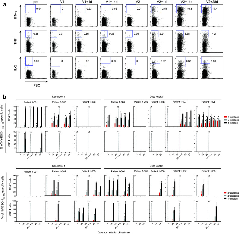
Vaccines that elicit targeted tumor antigen-specific T-cell responses have the potential to be used as adjuvant therapy in patients with high risk of relapse. However, the responses induced by vaccines in cancer patients have generally been disappointing. To improve vaccine function, we investigated the possibility of exploiting the immunostimulatory capacity of type 1 Natural killer T (NKT) cells, a cell type enriched in lymphoid tissues that can trigger improved antigen-presenting function in dendritic cells (DCs). In this phase I dose escalation study, we treated eight patients with high-risk surgically resected stage II-IV melanoma with intravenous autologous monocyte-derived DCs loaded with the NKT cell agonist α-GalCer and peptides derived from the cancer testis antigen NY-ESO-1. Two synthetic long peptides spanning defined immunogenic regions of the NY-ESO-1 sequence were used. This therapy proved to be safe and immunologically effective, inducing increases in circulating NY-ESO-1-specific T cells that could be detected directly ex vivo in seven out of eight patients. These responses were achieved using as few as 5 × 10⁵ peptide-loaded cells per dose. Analysis after in vitro restimulation showed increases in polyfunctional CD4+ and CD8+ T cells that were capable of manufacturing two or more cytokines simultaneously. Evidence of NKT cell proliferation and/or NKT cell-associated cytokine secretion was seen in most patients. In light of these strong responses, the concept of including NKT cell agonists in vaccine design requires further investigation.

Keywords: Dendritic cell; Melanoma; NKT cell; NY-ESO-1; α-Galactosylceramide.


Conflict of interest statement

The authors declare that they have no conflict of interest.

Figures

Fig. 1
Fig. 1
Study schema. Arrows indicate times when blood samples were collected for serum or PBMCs. Sample collections’ time-point is indicated in days (d) and hours (h)
Fig. 2
Fig. 2
Analysis of immunologic responses to vaccination. a Analysis of NKT cell frequency in patients over the course of the study. Points shown (open symbols) are the percentage of NKT cells in each of the duplicate samples at each sampled time-point, with mean values (closed symbols) shown as line graphs. Lines V1 and V2 indicate the two vaccination times. Statistically significant increases between post-vaccination measures and baseline (days − 2 and 0) are indicated by * for FWER corrected p < 0.05. b Analysis of NKT cell frequency in PBMCs ex vivo by IFN-γ ELISpot in response to α-GalCer. Points (open symbols) are numbers of IFN-γ spot forming units (SFC) in triplicate samples at each time-point, with mean values (closed symbols) plotted as line graphs. Statistically significant differences between post-vaccination time-points and baseline are indicated by * for FWER corrected p < 0.05. c Analysis of serum cytokines. Heat map indicates serum values in post-vaccination samples that were greater than two standard deviations above (pink) or below (blue) baselines levels. Upper panels show pre-specified cytokines that were above level of detection; lower panel show post hoc analysis of cytokines that were above level of detection. Statistically significant increases between post-vaccination measures and baseline are indicated by * for FWER corrected p < 0.05. Cytokines where this was greater than two standard deviations are marked red. d Upper panels show analysis of frequency of NY-ESO-1118–143-specific cells in PBMCs by ex vivo IFN-γ ELISpot. Points (open symbols) are numbers of SFC in triplicate samples at each time-point, with mean values (closed symbols) plotted as line graphs. Statistically significant increases between post-vaccination measures and baseline are indicated by * for FWER corrected p < 0.05. Middle and lower panels show flow cytometric analysis of IFN-γ+ cells after 10 days of restimulation with NY-ESO-1118–143 long peptide, with gating on CD4+ T cells (middle) or CD8+ T cells (lower). Points (open symbols) are percentages of IFN-γ+ cells in triplicate PBMC samples, with mean values (closed symbols) plotted as line graphs. Statistically significant increases between background-subtracted post-vaccination measures and baseline are indicated by * for FWER corrected p < 0.05. e As in d, except showing analysis of responses to NY-ESO-179–116 long peptide. f Flow cytometric analysis of IFN-γ+, TNF+ or IL-2+ cells after 10 days of restimulation on indicated long peptides, with gating on CD4+ or CD8+ T cells. Heat map indicates frequencies in post-vaccination samples that were greater than three standard deviations above pre-vaccination (pre) baselines levels. Statistically significant increases between post-vaccination measures and baseline are indicated by * for FWER corrected p < 0.05. Cytokines where this was greater than three standard deviations are marked red
Fig. 3
Fig. 3
Polyfunctionality analysis of NY-ESO-1-specific T cells after 10 day restimulation with NY-ESO-1 peptides. a Representative flow cytometry plots of NY-ESO-1118–143-reactive CD4+ T cells secreting IFN-γ, TNF and IL-2 from patient 1-007. b Percentages of cells expressing 1, 2, or 3 cytokines (functions) at once at the indicated time-points. Mean values ± SEM (standard error of the mean) for triplicates samples are shown for the indicated peptide-reactive cells. Statistically significant increases of bi- or tri-functional T-cell responses between post-vaccination measures and baseline are indicated by * for FWER corrected p < 0.05

References

    1. Kirkwood JM, Strawderman MH, Ernstoff MS, Smith TJ, Borden EC, Blum RH. Interferon alfa-2b adjuvant therapy of high-risk resected cutaneous melanoma: the Eastern Cooperative Oncology Group Trial EST 1684. J Clin Oncol. 1996;14(1):7–17. doi: 10.1200/JCO.1996.14.1.7. - DOI - PubMed
    1. Lens MB, Dawes M. Interferon alfa therapy for malignant melanoma: a systematic review of randomized controlled trials. J Clin Oncol. 2002;20(7):1818–1825. doi: 10.1200/JCO.2002.07.070. - DOI - PubMed
    1. Hodi FS, O’Day SJ, McDermott DF, Weber RW, Sosman JA, Haanen JB, Gonzalez R, Robert C, Schadendorf D, Hassel JC, Akerley W, van den Eertwegh AJ, Lutzky J, Lorigan P, Vaubel JM, Linette GP, Hogg D, Ottensmeier CH, Lebbe C, Peschel C, Quirt I, Clark JI, Wolchok JD, Weber JS, Tian J, Yellin MJ, Nichol GM, Hoos A, Urba WJ. Improved survival with ipilimumab in patients with metastatic melanoma. N Engl J Med. 2010;363(8):711–723. doi: 10.1056/NEJMoa1003466. - DOI - PMC - PubMed
    1. Larkin J, Chiarion-Sileni V, Gonzalez R, Grob JJ, Cowey CL, Lao CD, Schadendorf D, Dummer R, Smylie M, Rutkowski P, Ferrucci PF, Hill A, Wagstaff J, Carlino MS, Haanen JB, Maio M, Marquez-Rodas I, McArthur GA, Ascierto PA, Long GV, Callahan MK, Postow MA, Grossmann K, Sznol M, Dreno B, Bastholt L, Yang A, Rollin LM, Horak C, Hodi FS, Wolchok JD. Combined nivolumab and ipilimumab or monotherapy in untreated melanoma. N Engl J Med. 2015;373(1):23–34. doi: 10.1056/NEJMoa1504030. - DOI - PMC - PubMed
    1. Robert C, Long GV, Brady B, Dutriaux C, Maio M, Mortier L, Hassel JC, Rutkowski P, McNeil C, Kalinka-Warzocha E, Savage KJ, Hernberg MM, Lebbe C, Charles J, Mihalcioiu C, Chiarion-Sileni V, Mauch C, Cognetti F, Arance A, Schmidt H, Schadendorf D, Gogas H, Lundgren-Eriksson L, Horak C, Sharkey B, Waxman IM, Atkinson V, Ascierto PA. Nivolumab in previously untreated melanoma without BRAF mutation. N Engl J Med. 2015;372(4):320–330. doi: 10.1056/NEJMoa1412082. - DOI - PubMed

Publication types

MeSH terms