. 2018 Apr;223(3):1133-1148.
doi: 10.1007/s00429-017-1542-8. Epub 2017 Nov 1.

Perineuronal nets labeled by monoclonal antibody VC1.1 ensheath interneurons expressing parvalbumin and calbindin in the rat amygdala

Affiliations

Perineuronal nets labeled by monoclonal antibody VC1.1 ensheath interneurons expressing parvalbumin and calbindin in the rat amygdala

Alexander J McDonald et al. Brain Struct Funct. 2018 Apr.

Abstract

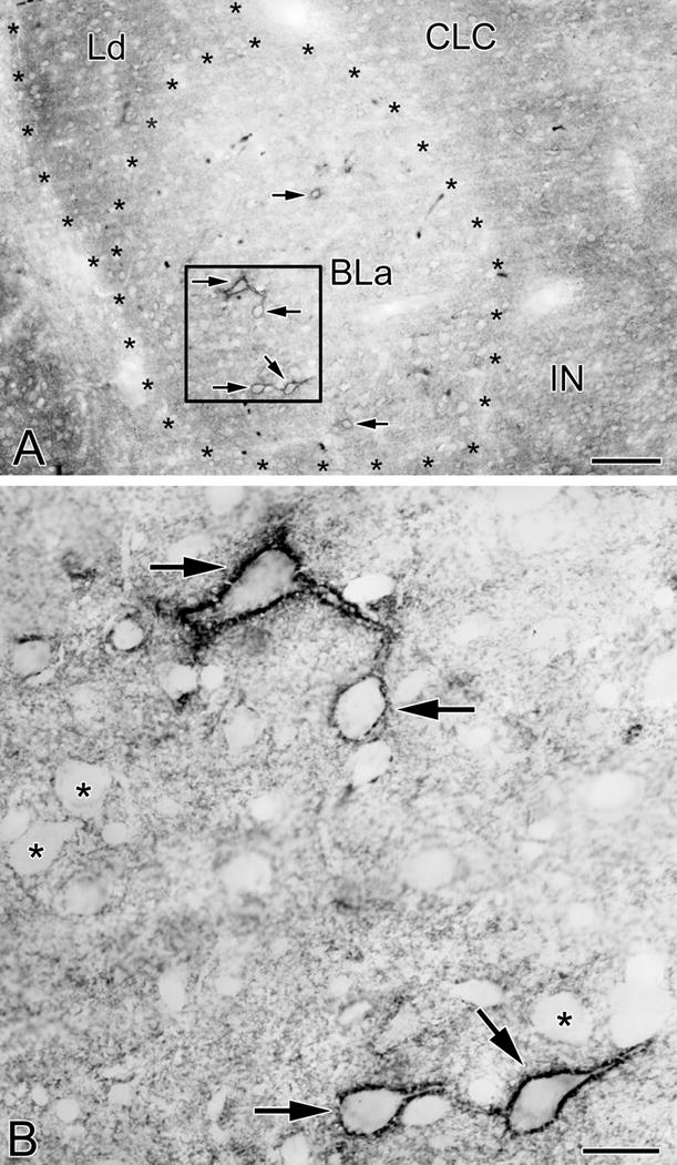
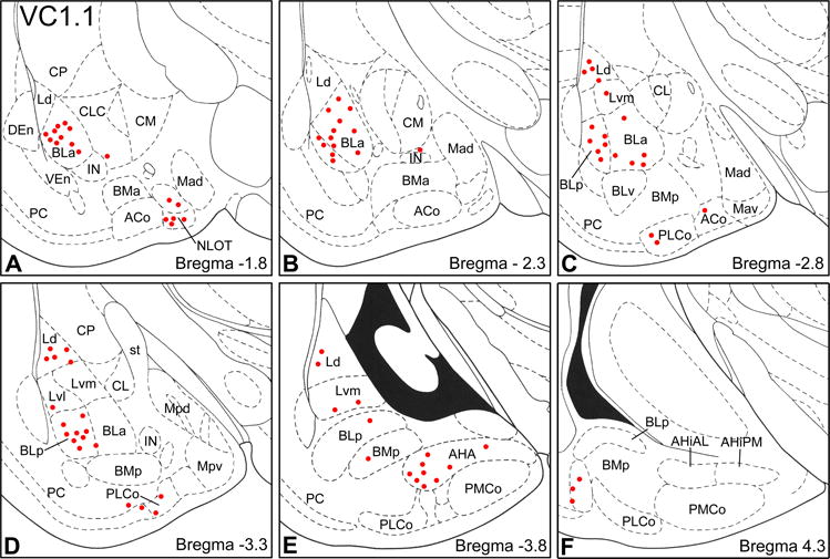
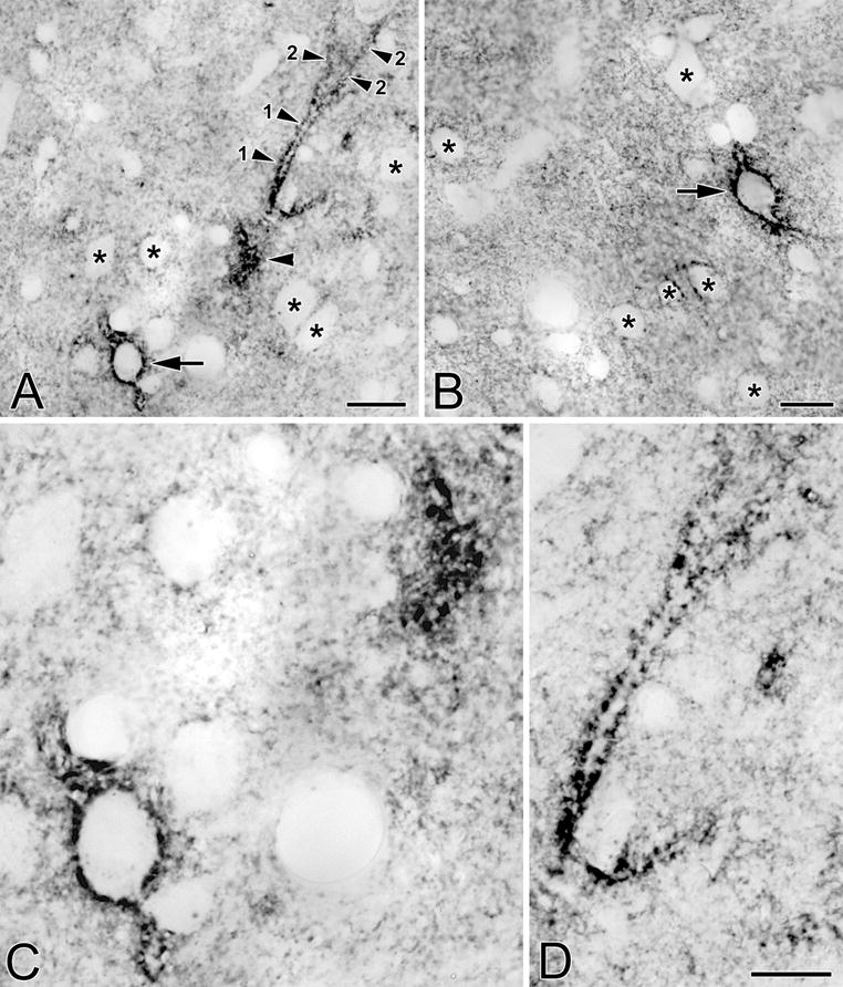
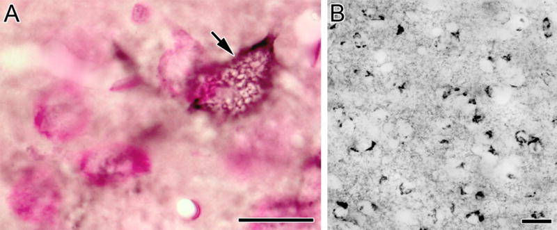
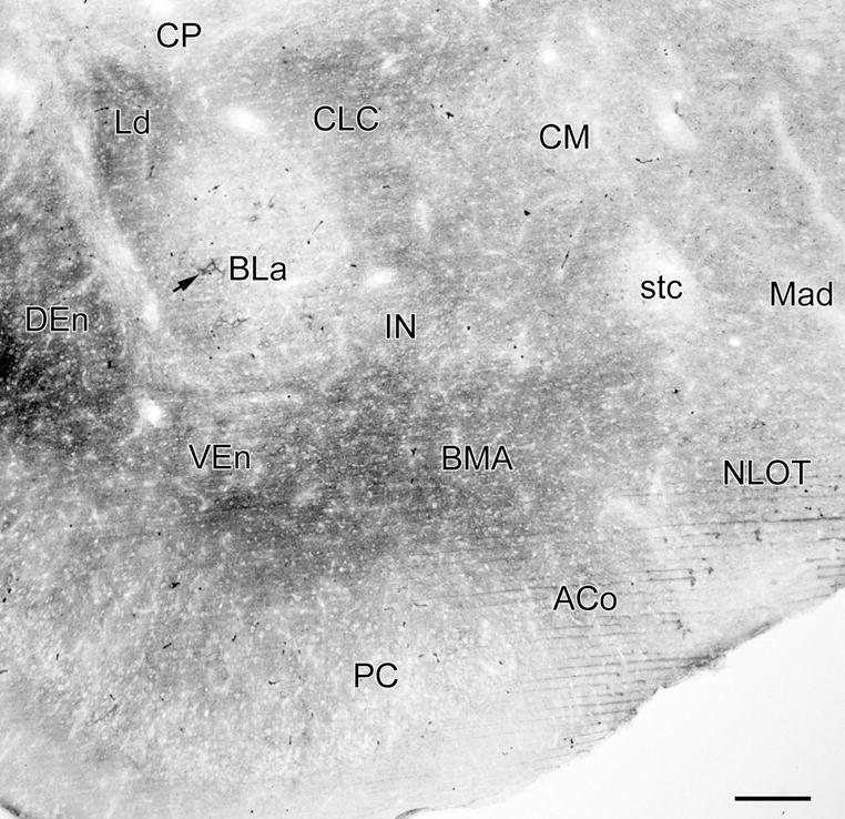
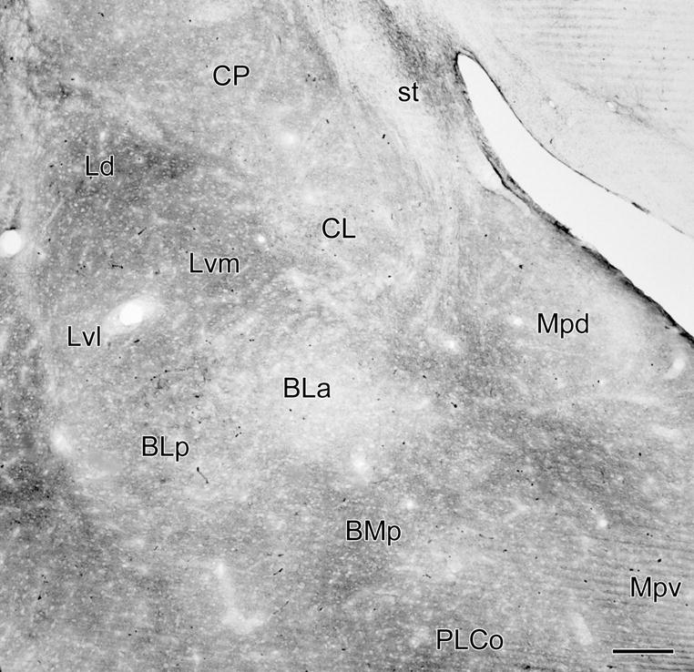
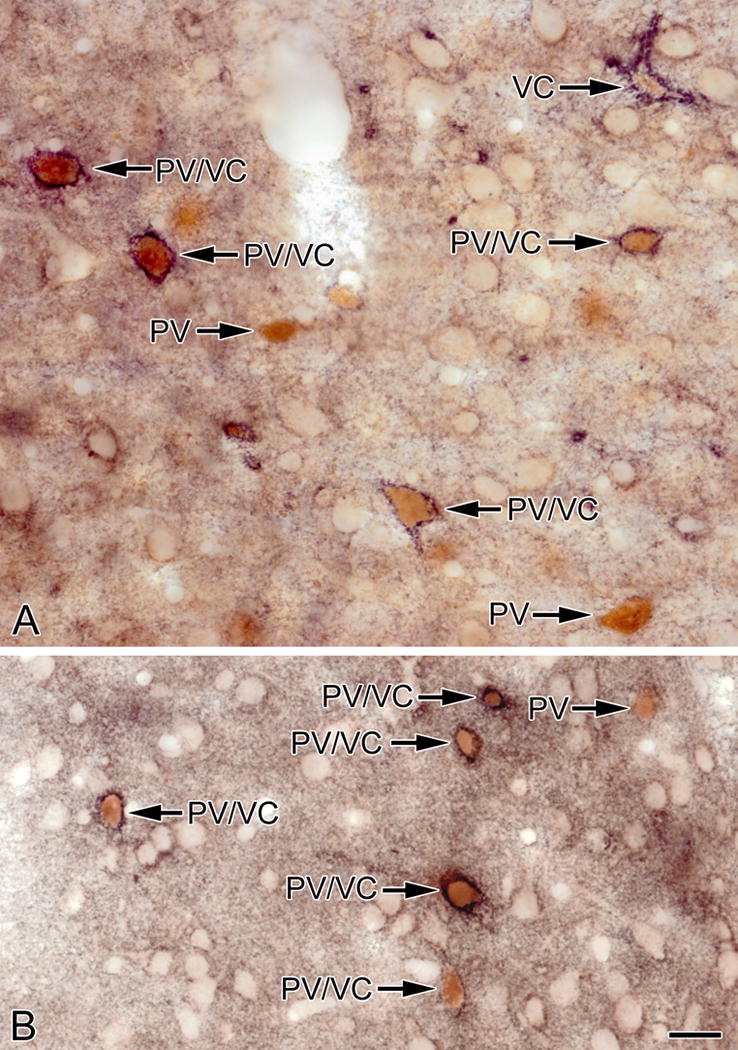
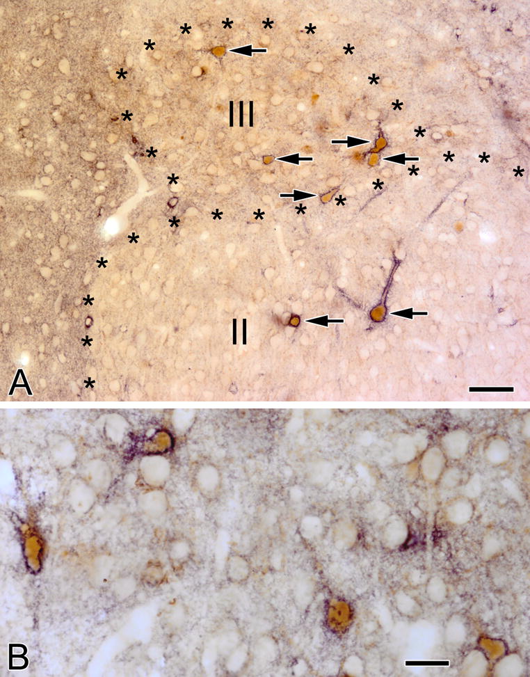
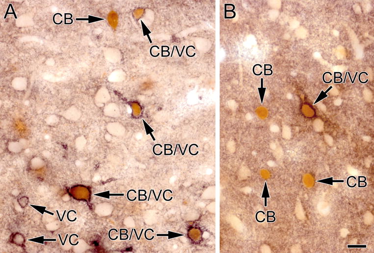
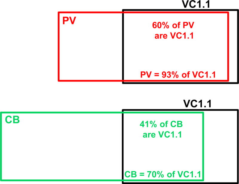
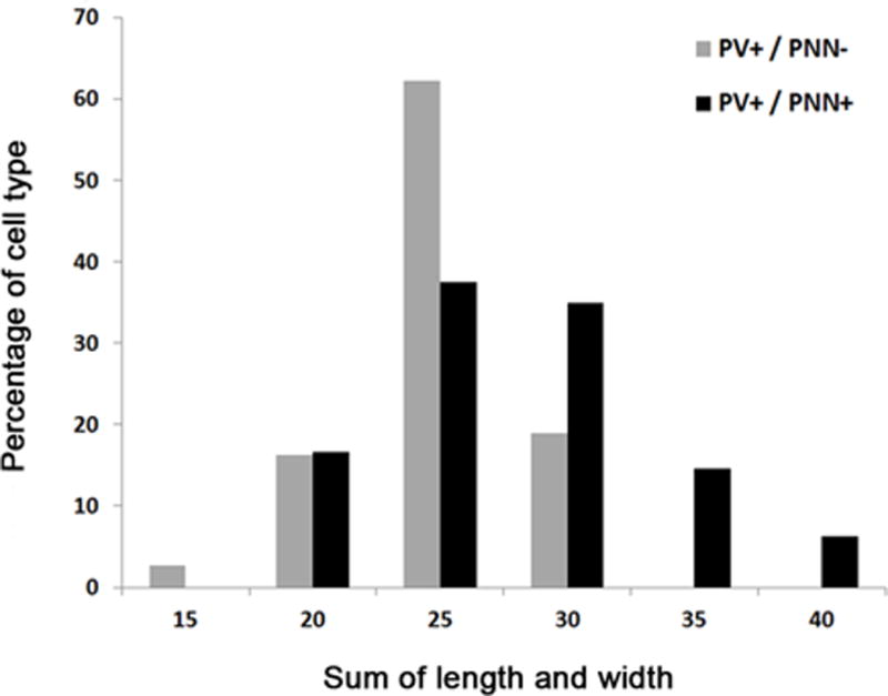
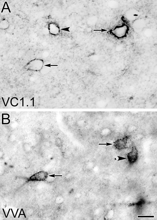
Perineuronal nets (PNNs) are specialized condensations of extracellular matrix that ensheath particular neuronal subpopulations in the brain and spinal cord. PNNs regulate synaptic plasticity, including the encoding of fear memories by the amygdala. The present immunohistochemical investigation studied PNN structure and distribution, as well as the neurochemistry of their ensheathed neurons, in the rat amygdala using monoclonal antibody VC1.1, which recognizes a glucuronic acid 3-sulfate glycan associated with PNNs in the cerebral cortex. VC1.1+ PNNs surrounded the cell bodies and dendrites of a subset of nonpyramidal neurons in cortex-like portions of the amygdala (basolateral amygdalar complex, cortical nuclei, nucleus of the lateral olfactory tract, and amygdalohippocampal region). There was also significant neuropilar VC1.1 immunoreactivity, whose density varied in different amygdalar nuclei. Cell counts in the basolateral nucleus revealed that virtually all neurons ensheathed by VC1.1+ PNNs were parvalbumin-positive (PV+) interneurons, and these VC1.1+/PV+ cells constituted 60% of all PV+ interneurons, including all of the larger PV+ neurons. Approximately 70% of VC1.1+ neurons were calbindin-positive (CB+), and these VC1.1+/CB+ cells constituted about 40% of all CB+ neurons. Colocalization of VC1.1 with Vicia villosa agglutinin (VVA) binding, which stains terminal N-acetylgalactosamines, revealed that VC1.1+ PNNs were largely a subset of VVA+ PNNs. This investigation provides baseline data regarding PNNs in the rat which should be useful for future studies of their function in this species.

Keywords: Amygdala; Calcium-binding proteins; Extracellular matrix; Immunohistochemistry; Perineuronal nets.


Conflict of interest statement

Conflict of interest: The authors declare that they have no conflict of interest.

Figures

Fig. 1
Fig. 1
VC1.1-ir in PNNs and the neuropil in the anterior subdivision of the basolateral nucleus (BLa). (A) Photomicrograph of VC1.1-ir in the BLa at the bregma −1.8 level. Asterisks indicate the borders of BLa and the dorsolateral subdivision of the lateral nucleus (Ld). Four neurons ensheathed by VC1.1+ PNNs are in the boxed area of BLa (arrows; see B). Two additional BLa neurons ensheathed by VC1.1+ PNNs are also indicated by arrows. Other abbreviations: CLC, lateral capsular subdivision of the central nucleus; IN, intercalated nucleus. (B) Higher power photomicrograph of the boxed area in A. Note punctate VC1.1-ir in the neuropil and along the plasma membranes of the cell bodies and proximal dendrites of four neurons (arrows). In addition, some VC1.1-ir in the neuropil is in the form of small rings that are 1-2 μm in diameter. Asterisks indicate representative neurons that are not ensheathed by PNNs, but whose plasma membranes are decorated by punctate neuropilar VC1.1-ir. Scale bars = 100 μm in A, 20 μm in B
Fig. 2
Fig. 2
Distribution of VC1.1+ PNNs in the amygdala (red dots) in sections arranged from rostral (A) to caudal (F). Each bregma level shows the locations of neurons plotted from three non-adjacent 50 μm-thick sections; each dot represents one neuron. Templates are modified from the atlas by Paxinos and Watson (1997). Abbreviations: ACo, anterior cortical nucleus; AHA, amygdalohippocampal area; AHiAL, anterolateral amygdalohippocampal area; AHiPM, posteromedial amygdalohippocampal area; BLa, anterior basolateral nucleus; BLp, posterior basolateral nucleus; BLv, ventral basolateral nucleus; BMa, anterior basomedial nucleus; BMp, posterior basomedial nucleus; CL, lateral central nucleus; CLC, lateral capsular subdivision of the central nucleus; CM, medial central nucleus; CP, caudate putamen; DEn, dorsal endopiriform nucleus; IN, intercalated nucleus; Ld, dorsolateral lateral nucleus; Lvl, ventrolateral lateral nucleus; Lvm, ventromedial lateral nucleus; Mad, anterodorsal medial nucleus; Mav, anteroventral medial nucleus; Mpd, posterodorsal medial nucleus; Mpv, posteroventral medial nucleus; NLOT, nucleus of the lateral olfactory tract; PC, piriform cortex; PLCo, posterolateral cortical nucleus; PMCo, posteromedial cortical nucleus; st, stria terminalis; VEn, ventral endopiriform nucleus.
Fig. 3
Fig. 3
(A) Photomicrograph of a neuron in the BLa that is ensheathed by a VC1.1+ PNN (arrow). Also in the field is a primary dendrite (1) and two secondary dendrites (2) of another neuron. Only the edge of the cell body giving rise to these dendrites was in this section (unlabeled arrowhead). (B) Photomicrograph of a neuron in the lateral nucleus that is ensheathed by a VC1.1+ PNN (arrow). (C) Higher power view of the cell bodies in A. (D) Higher power view of the dendrites in A. Asterisks in A, B and C indicate representative neurons that are not ensheathed by PNNs, but whose plasma membranes are decorated by punctate neuropilar VC1.1-ir. Scale bars = 20 μm for A and B; 10 μm in D (C is at the same magnification)
Fig. 4
Fig. 4
(A) Photomicrograph of a VC1.1+ PNN (black) surrounding the cell body of a BLa neuron (arrow) in a pyronin Y counterstained section. This en face view of the VC1.1+ lattice coating the plasma membrane of this cell shows ovoid gaps that probably correspond to locations where axon terminals are forming synaptic contacts with this cell body. (B) VC1.1-ir in the lateral subdivision of the central nucleus. Scale bars = 20 μm
Fig. 5
Fig. 5
Photomicrograph of VC1.1-ir in a coronal section through the rostral amygdala (bregma −1.8 level; compare with Fig. 2A; see caption of Fig. 2 for abbreviations). Figure 1A is a higher power photomicrograph of the BLa in this section. Arrow indicates a large neuron in the anterior subdivision of the basolateral nucleus (BLa) that is ensheathed by a VC1.1+ PNN. There are 5 additional PNNs in the BLa in this section, and 3 in the nucleus of the lateral olfactory tract (NLOT), but they are difficult to see at this low magnification. Thus, almost all of the VC1.1-ir seen in this section is in the neuropil. Scale bar = 200 μm
Fig. 6
Fig. 6
Photomicrograph of VC1.1-ir in a coronal section through the middle of the amygdala (bregma −3.3 level; compare with Fig. 2D). Almost all of the VC1.1-ir seen in this section is in the neuropil. Scale bar = 200 μm
Fig. 7
Fig. 7
Photomicrographs of sections through the BLa (A) and lateral nucleus (B) stained for VC1.1 (black) and PV (brown). Most PV+ neurons are ensheathed by VC1.1+ PNNs (PV/VC) but some are not (PV). One neuron in A is ensheathed by a VC1.1+ PNN but exhibits little or no PV immunoreactivity (VC). Scale bar = 20 μm for both A and B
Fig. 8
Fig. 8
(A) VC1.1+ PNNs (black) surround PV+ neurons (brown) in the nucleus of the lateral olfactory tract. Two of these neurons are in layer II and five are in layer III (arrows). (B) Four PV+ neurons (brown) in the amygdalohippocampal area are ensheathed by VC1.1+ PNNs (black). Scale bar = 50 μm in A and 20 μm in B
Fig. 9
Fig. 9
Photomicrographs of sections through the BLa (A) and lateral nucleus (B) stained for VC1.1 (black) and CB (brown). Several CB+ neurons are ensheathed by VC1.1+ PNNs (CB/VC) but some are not (CB). Two neurons in A are ensheathed by a VC1.1+ PNN but exhibit no CB immunoreactivity (VC). Scale bar = 20 μm for both A and B
Fig. 10
Fig. 10
Venn diagrams illustrating the relationship of PV+ neurons (upper diagram) and CB+ neurons (lower diagram) to VC1.1+ neurons in the BLa. The sizes of the rectangles indicate the relative numbers of the different neuronal populations. Note that CB+ neurons outnumber PV+ neurons in the BLa. Approximately 80% of PV+ neurons are CB+, and these PV+/CB+ neurons constitute about 60% of the CB+ population (McDonald and Betette, 2001)
Fig. 11
Fig. 11
Histogram showing the perikaryal sizes of PV+ interneurons ensheathed by VC1.1+ PNNs (PV+/PNN+; black) and PV+ interneurons not ensheathed by VC1.1+ PNNs (PV+/PNN–; gray) in the BLa
Fig. 12
Fig. 12
Photomicrographs of corresponding surfaces of adjacent sections through the BLa stained with the VC1.1 antibody (A) or VVA (B). Arrows indicate two VC1.1+/VVA+ neurons that were stained in both sections. Arrowhead in A indicates a single-labeled VC1.1+ neuron. Arrowhead in B indicates a single-labeled VVA+ neuron. Scale bar = 20 μm for both A and B
Fig. 13
Fig. 13
Distribution of VVA+ PNNs in the amygdala (red dots) in sections arranged from rostral (A) to caudal (F). Each bregma level shows the locations of neurons plotted from three non-adjacent 50 μm-thick sections; each dot represents one neuron. Templates are modified from the atlas by Paxinos and Watson (1997). For abbreviations see caption for Figure 2.

References

    1. Abo T, Balch CM. A differentiation antigen of human NK and K cells identified by a monoclonal antibody (HNK-1) J Immunol. 1981;127:1024–1029. - PubMed
    1. Alpár A, Gärtner U, Härtig W, Brückner G. Distribution of pyramidal cells associated with perineuronal nets in the neocortex of rat. Brain Res. 2006;1120:13–22. - PubMed
    1. Arimatsu Y, Naegele JR, Barnstable CJ. Molecular markers of neuronal subpopulations in layers 4, 5, and 6 of cat primary visual cortex. J Neurosci. 1987;7:1250–1263. - PMC - PubMed
    1. Baker KD, Gray AR, Richardson R. The development of perineuronal nets around parvalbumin gabaergic neurons in the medial prefrontal cortex and basolateral amygdala of rats. Behav Neurosci. 2017;131:289–303. - PubMed
    1. Balmer TS. Perineuronal Nets Enhance the Excitability of Fast-Spiking Neurons. eNeuro. 2016 Jul 27;3(4) - PMC - PubMed

LinkOut - more resources