

Clinical Implications of Pediatric Colonic Eosinophilia
- PMID: 29095349
- PMCID: PMC5916023
- DOI: 10.1097/MPG.0000000000001784
Clinical Implications of Pediatric Colonic Eosinophilia
Abstract
Objective: Pediatric colonic eosinophilia represents a confounding finding with a wide differential. It is often difficult to determine which children may progress to inflammatory bowel disease (IBD), which have an eosinophilic colitis (EC), and which may have no underlying pathology. There is little guidance for the practitioner on the approach to these patients. To define the clinical presentations of colonic eosinophilia and identify factors which may aid in diagnosis we reviewed patients with colonic eosinophilia and the clinicopathologic factors associated with their diagnoses.
Methods: An 8-year retrospective chart review of children whose histopathology identified colonic eosinophilia (N = 72) compared to controls with normal biopsies (N = 35).
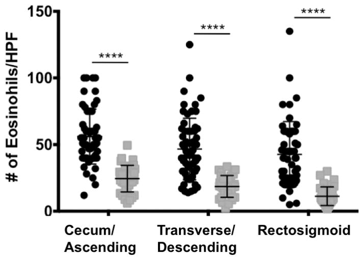
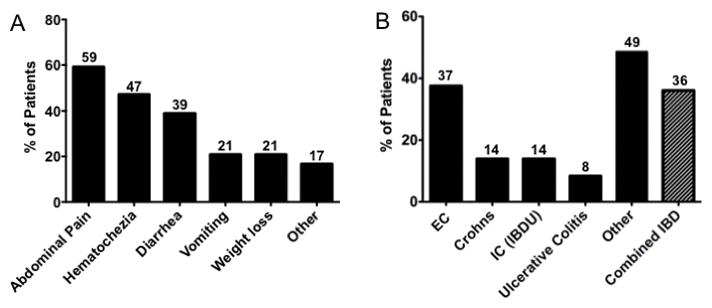
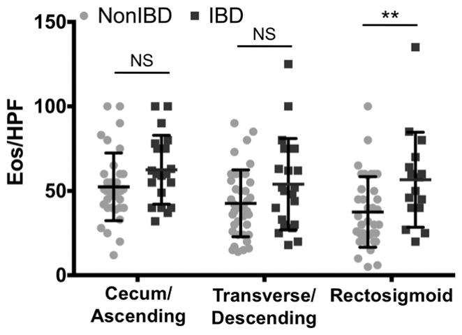
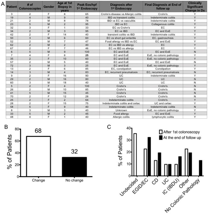
Results: Patients with colonic eosinophilia had increased eosinophils/high-power field compared to controls (P < 0.001) and had 3 clinical phenotypes. Thirty-six percent had an inflammatory phenotype with elevated erythrocyte sedimentation rate (P < .0001), chronic inflammation on colonic biopsies (P < 0.001), and were diagnosed as having IBD. Thirty-seven percent were diagnosed as having EC, associated with male sex (P < 0.005) and peripheral eosinophilia (P = 0.041). Twenty-one percent had no significant colonic pathology. Forty-three percent of patients had >1 colonoscopy and 68% of these had change from initial diagnoses.
Conclusions: There are 3 main phenotypes of children with colonic eosinophilia. Signs of chronic systemic inflammation raise suspicion for IBD. Peripheral eosinophilia and male sex are associated with EC. A significant percent of children with colonic eosinophilia do not have colonic disease. Eosinophils/high-power field is not reliable to differentiate etiologies. Repeat colonoscopies may be required to reach final diagnoses.
Figures
References
-
- Saad AG. Normal quantity and distribution of mast cells and eosinophils in the pediatric colon. Pediatr Dev Pathol. 2011;14:294–300. - PubMed
-
- DeBrosse CW, Case JW, Putnam PE, et al. Quantity and distribution of eosinophils in the gastrointestinal tract of children. Pediatr Dev Pathol. 2006;9:210–8. - PubMed
-
- Lowichik A, Weinberg AG. A quantitative evaluation of mucosal eosinophils in the pediatric gastrointestinal tract. Mod Pathol. 1996;9:110–4. - PubMed
-
- Chernetsova E, Sullivan K, de Nanassy J, et al. Histologic analysis of eosinophils and mast cells of the gastrointestinal tract in healthy Canadian children. Hum Pathol. 2016;54:55–63. - PubMed
-
- Pascal RR, Gramlich TL, Parker KM, et al. Geographic variations in eosinophil concentration in normal colonic mucosa. Modern pathology : an official journal of the United States and Canadian Academy of Pathology, Inc. 1997;10:363–365. - PubMed
Publication types
MeSH terms
Grants and funding
LinkOut - more resources
Full Text Sources
Other Literature Sources
Medical
