Multicenter Study
. 2018 Jan;38 Suppl 1(Suppl 1):S43-S50.
doi: 10.1097/IAE.0000000000001842.

CHARACTERISTICS OF PIGMENTED LESIONS IN TYPE 2 IDIOPATHIC MACULAR TELANGIECTASIA

Affiliations
Multicenter Study

CHARACTERISTICS OF PIGMENTED LESIONS IN TYPE 2 IDIOPATHIC MACULAR TELANGIECTASIA

Irene Leung et al. Retina. 2018 Jan.

Abstract

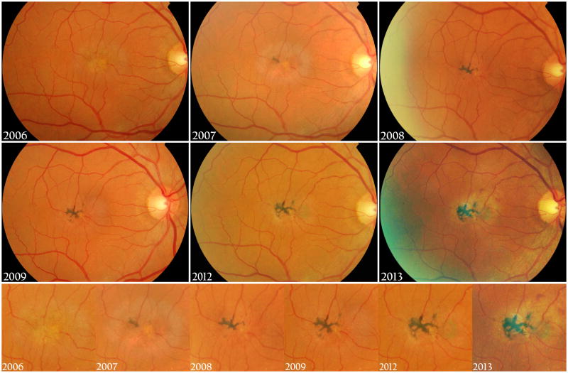
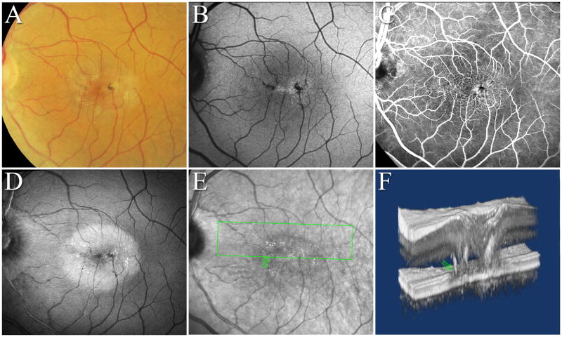
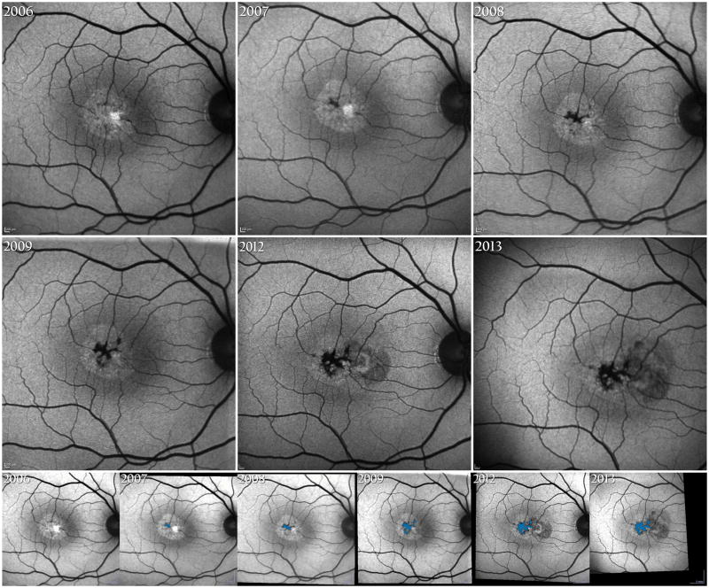
Purpose: Pigment in the midretina is a characteristic sign in Type 2 idiopathic macular telangiectasia (MacTel) and is considered to characterize the late stage of the disease. Our aim was to investigate its incidence, and relationship with risk factors for MacTel, including outer retinal vascularization and subretinal neovascular proliferation (SRNV).

Methods: Pigment extent was measured in fundus autofluorescence images of 150 eyes of 75 MacTel probands, using the Region Finder tool of Heidelberg Eye Explorer. A linear mixed model was used to analyze the dynamics of pigment and its associations with other features of the phenotype. The relative incidence of pigment and of outer retinal outer retinal vascularization and SRNV was analyzed within the full MacTel Study cohort (1,244 probands).

Results: Mean pigment area at baseline was 0.157 mm (range = 0-1.295 mm, SD = 0.228 mm, n = 101). Progression demonstrated a nonlinear pattern (P < 0.001) at an overall rate of 0.0177 mm/year and was associated with the initial plaque size and with SRNV. There was a strong correlation between fellow eyes (P ≤ 0.0001). In approximately 25% of all SRNV cases, SRNV may coincide with or precede pigment.

Conclusion: Our data may be useful for refining the current system for staging disease severity in MacTel.


Figures

Figure 1
Figure 1. Progression of abnormal pigment in MacTel in color fundus images
Typically, brown (melanin-like) pigment first appears focally temporal of the foveal centre, involving abnormal outer retinal blood vessels. Over time it may extend to the foveal centre. Clearly visible pigment plaques are typically located to the mid-retina.
Figure 2
Figure 2. Typical MacTel pigment plaques in multiple imaging modalities
A. CF, B. autofluorescence, C. early FFA, D. blue light reflectance image, E. infrared and F.: 3D perspective view of OCT volume data. As the OCT perspective view illustrates, the intraretinal pigment is located surrounding abnormal blood vessels originating in the deep retinal plexus and traversing the retinal layers towards the RPE (matching green arrows). Most of the pigment in this eye is located external to the superficial and deep retinal meshworks, the apparent extent of the pigment is much smaller in the FFA image than in the FAF and CF images. The source of the pigment is hypothesized to be inside cells migrating from the RPE.
Figure 3
Figure 3. Top and Middle rows: Corresponding SLO AF images of the eye presented in Figure 1
Increased central AF due to loss of luteal pigment is apparent. Decreased AF (intense irregular dark areas) is caused by the masking effect of pigment in the mid-retina, involving abnormal blood vessels. This needs to be differentiated from dense scar tissue, blood or subretinal neovascular proliferation (2012-). Bottom row shows the pigment area progression captured by the Region Finder tool of Heidelberg Eye Explorer (blue area).

References

    1. Gass JD, Oyakawa RT. Idiopathic juxtafoveolar retinal telangiectasis. Arch Ophthalmol. 1982;100:769–780. - PubMed
    1. Gass JD, Blodi BA. Idiopathic juxtafoveolar retinal telangiectasis. Update of classification and follow-up study. Ophthalmology. 1993;100:1536–1546. - PubMed
    1. Charbel Issa P, Gillies MC, Chew EY, et al. Macular telangiectasia type 2. Prog Retin Eye Res. 2013;34:49–77. - PMC - PubMed
    1. Zhao M, Andrieu-Soler C, Kowalczuk L, et al. A new CRB1 rat mutation links Muller glial cells to retinal telangiectasia. J Neurosci. 2015;35:6093–6106. - PMC - PubMed
    1. Dorrell MI, Aguilar E, Jacobson R, et al. Antioxidant or neurotrophic factor treatment preserves function in a mouse model of neovascularization-associated oxidative stress. J Clin Invest. 2009;119:611–623. - PMC - PubMed

Publication types

MeSH terms

Substances

Supplementary concepts