

WIDE-FIELD SMARTPHONE FUNDUS VIDEO CAMERA BASED ON MINIATURIZED INDIRECT OPHTHALMOSCOPY
- PMID: 29095361
- PMCID: PMC5777875
- DOI: 10.1097/IAE.0000000000001888
WIDE-FIELD SMARTPHONE FUNDUS VIDEO CAMERA BASED ON MINIATURIZED INDIRECT OPHTHALMOSCOPY
Abstract
Purpose: This study is to develop a low-cost, easy-to-use, wide-field smartphone fundus video camera to enable affordable point-of-care examination and telemedicine.
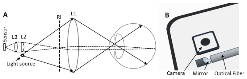
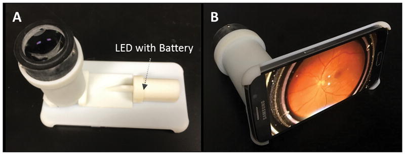
Methods: The wide-field smartphone fundus camera is based on a unique design of miniaturized indirect ophthalmoscopy. For proof-of-concept prototype, we used a Samsung Galaxy S6 smartphone and all off-the-shelf components. A fiber coupled LED was used to deliver illumination light through a 1 mm micro mirror, which was conjugated to the subject pupil plane, for retinal illumination. A 60D ophthalmic lens was used to image the retina through the eye, and a plano-convex lens with 90 mm focal length was used to relay the retinal image to the smartphone camera.
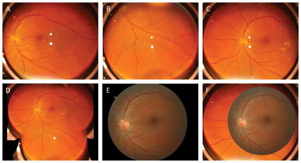
Results: A totally wireless smartphone fundus camera was constructed, with a whole weight of 255 g. This device allowed both snapshot fundus photography and continuous video recording. 92° field of view (FOV) was achieved in single-shot images. Optic disc, macula, and retinal blood vasculatures can be clearly observed with image quality comparable to standard fundus camera.
Conclusion: Miniaturized indirect ophthalmoscopy enabled a low-cost, portable, wide-field smartphone fundus camera, which can foster telemedicine and clinical deployments of wide-field fundus photography for eye disease screening, diagnosis and treatment assessment.
Figures
References
-
- Maamari RN, Keenan JD, Fletcher DA, Margolis TP. A mobile phone-based retinal camera for portable wide field imaging. The British journal of ophthalmology. 2014;98:438–441. - PubMed
-
- Suto S, Hiraoka T, Oshika T. Fluorescein fundus angiography with smartphone. Retina (Philadelphia, Pa) 2014;34:203–205. - PubMed
Publication types
MeSH terms
Grants and funding
LinkOut - more resources
Full Text Sources
Other Literature Sources
Medical
