. 2017 Nov 2;12(11):e0187476.
doi: 10.1371/journal.pone.0187476. eCollection 2017.

Concurrent jellyfish blooms and tenacibaculosis outbreaks in Northern Norwegian Atlantic salmon (Salmo salar) farms

Affiliations

Concurrent jellyfish blooms and tenacibaculosis outbreaks in Northern Norwegian Atlantic salmon (Salmo salar) farms

Sverre Bang Småge et al. PLoS One. .

Erratum in

Abstract

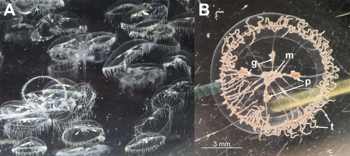
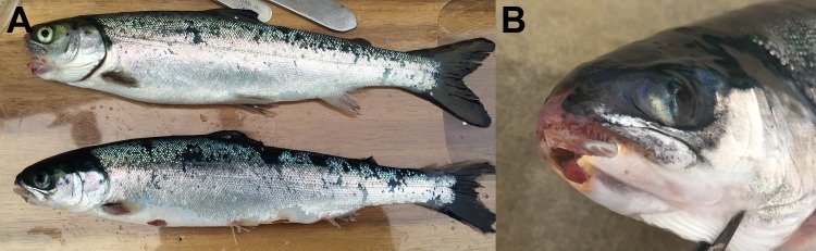
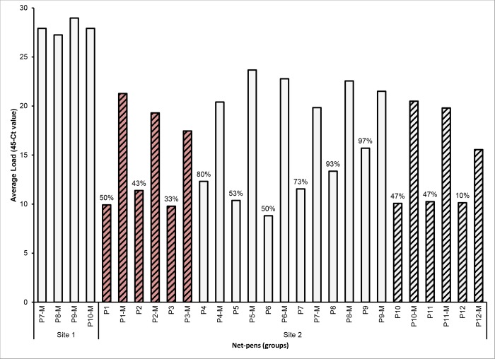
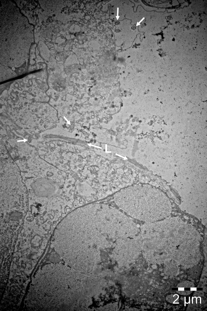
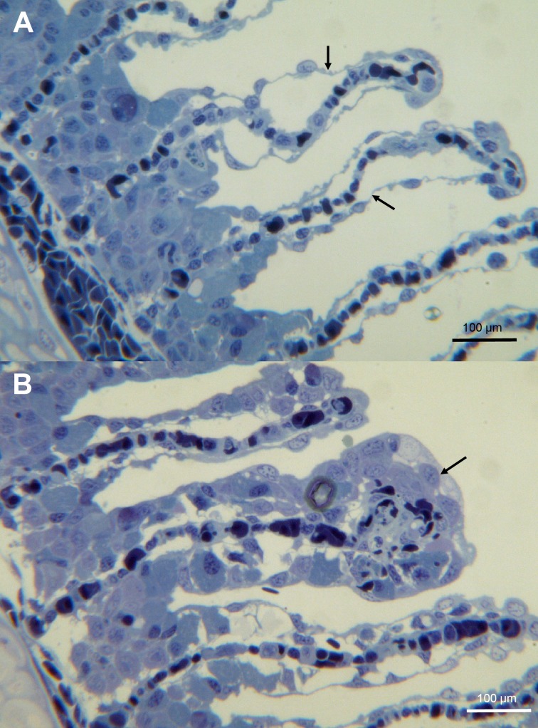
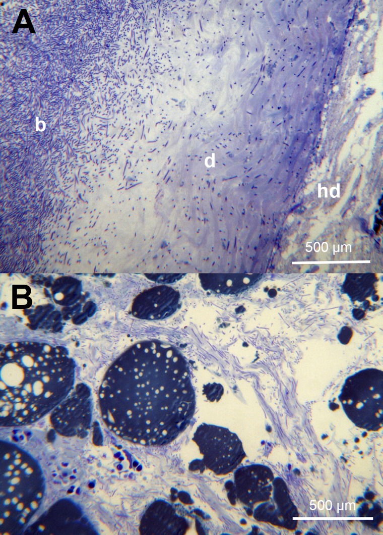
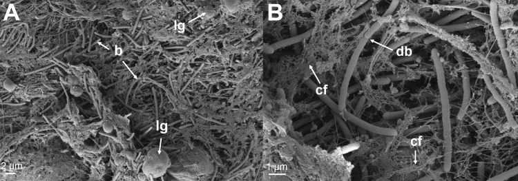
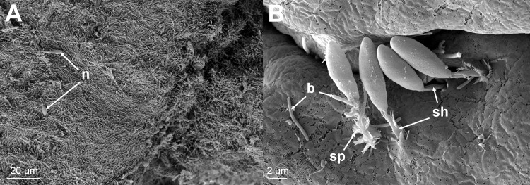
Tenacibaculosis is an increasing problem in the Norwegian Atlantic salmon aquaculture industry causing significant economic losses. In September 2015, two separate outbreaks of suspected tenacibaculosis occurred at two Atlantic salmon farms in Finnmark County in Northern Norway. The events resulted in major losses of smolts newly transferred into seawater. Prior to, and during the outbreaks, large numbers of small jellyfish, identified as Dipleurosoma typicum (Boeck) were observed in the vicinity of the farms and inside the net-pens. This study investigates the possible link between the jellyfish, Tenacibaculum spp. and the tenacibaculosis outbreaks. Bacteriology, histology, scanning and transmission electron microscopy, and real-time RT-PCR screening were performed on both fish and jellyfish samples. Based on the findings, Tenacibaculum finnmarkense was found to be the dominant bacteria associated with the tenacibaculosis outbreaks at both sites and that D. typicum is unlikely to be a vector for this fish pathogenic bacterium. However, results do show that the jellyfish caused direct damage to the fish's skin and may have exacerbated the bacterial infection by allowing an entry point for bacteria.


Conflict of interest statement

Competing Interests: The authors have declared that no competing interests exist.

Figures

Fig 1
Fig 1. Map of sampling points.
Map showing the area surrounding Porsangerhalvøya in Finnmark in the northernmost part of Norway. Farm sites are marked as S1 and S2 and environmental sampling points are marked with the site number, followed by the environmental sampling point number (e.g. S1E2).
Fig 2
Fig 2. Jellyfish morphology.
(A) A water sample collected outside one of the net-pens at site 1. A high concentration of D. typicum was present in the sample. (B) One of these specimens under a stereomicroscope showing the characteristics typical for this species [40,41]. m: the mouth, t: tentacles, g: gonads, p: planula larva (Note: the planula larva was found to be ciliated and highly motile, which differs from previous descriptions given for this species).
Fig 3
Fig 3. Moribund fish showing signs of tenacibaculosis.
(A) Fish representing moribunds from both site 1 and 2. Skin lesions have the typical scale loss and yellow margins seen with tenacibaculosis. Frayed fins are also clearly visible. (B) A close up of an affected head showing a mouth lesion that has the typical yellow margin associated with tenacibaculosis.
Fig 4
Fig 4. Phylogenetic analysis of recovered Tenacibaculum spp. isolates.
Phylogenetic tree of the 16S rRNA gene sequences obtained in this study and all known type strains in genus Tenacibaculum. In addition, Flexibacter echinicida and Flexibacter aurantiacus subsp. copepodarumT were included. A Bayesian analysis was performed using the GTR+G+I model.
Fig 5
Fig 5. Real-time RT-PCR results from sampled fish.
Average load of Tenacibaculum spp. positive fish for each sampled group measured with the Tb tuf real-time RT-PCR assay. Dataset includes both randomly sampled fish from each pen at site 2 (n = 30) and moribunds (-M) from each affected pen (n = 10 for site 1 and n = 5 for site 2). The prevalence in the moribunds was 100% and the prevalence in the randomly sampled fish is indicated as % above the respective bar. Striped columns are spring smolts and non-striped columns are autumn smolts. Red columns were treated with florfenicol prior to sampling.
Fig 6
Fig 6. TEM micrograph of the mouth region of D. typicum.
The typical 9+2 pattern (arrows) of motile ciliary axonemes are clearly visible. A cilium can be seen longitudinally (L).
Fig 7
Fig 7. SEM micrographs of D. typicum mouth region.
(A) A SEM micrograph showing the lip of the mouth of D. typicum. (B) A higher magnification of the inside of the mouth showing an abundance of cilia (c) and spherical structures that are likely droplets of exoenzymes (de) important in the digestion of food [10].
Fig 8
Fig 8. Histology of gills from moribund fish.
(A) A toluidine blue stained section of gills showing epithelial lifting (arrow). (B) A toluidine blue stained section of gills showing foci of hypertrophy (arrow).
Fig 9
Fig 9. Histology of skin from moribund fish.
(A) A toluidine blue stained section of skin showing complete loss of epidermis where the bacteria (b) are present and infiltration of the stratum compactum of the dermis (d). This section does not appear to have bacteria infiltrating the hypodermis (hd). (B) A toluidine blue stained section of the skin hypodermis showing large number of bacteria.
Fig 10
Fig 10. SEM micrographs of lesions from moribund fish.
(A) A SEM micrograph from the margin of a jaw lesion showing Tenacibaculum-like bacteria (b) infiltrating the dermis layer of the skin. Lipids globules (lg) can be seen and are likely a result of the destruction of the dermis layer as seen histologically. (B) A higher magnification showing a Tenacibaculum-like bacterial cell replicating in the dermis (db). The breakdown of the collagen fibers (cf) of the dermis can be seen associated with the bacteria.
Fig 11
Fig 11. SEM micrographs.
(A) A SEM micrograph from the margin of a lesion showing a mat of Tenacibaculum-like bacteria with a few attached nematocysts (n). (B) A higher magnification of one of these areas showing the nematocysts with their spiked (sp) covered shafts (sh) embedded in the skin. Bacteria (b) can be seen in close proximity with some seeming to be entering the holes created by the nematocysts.

References

    1. Rutter T. Managing risk and uncertainty: OECD, Paris; 2010 [updated 15-16th April]. Available from: http://www.oecd.org/greengrowth/fisheries/45400772.pdf.
    1. Rodger HD, Henry L, Mitchell SO. Non-infectious gill disorders of marine salmonid fish. Reviews in Fish Biology and Fisheries. 2011;21(3):423–40. doi: 10.1007/s11160-010-9182-6 - DOI
    1. Boero F, Bouillon J, Gravili C, Miglietta MP, Parsons T, Piraino S. Gelatinous plankton: irregularities rule the world (sometimes). Marine Ecology Progress Series. 2008;356:299–310.
    1. Purcell JE, Uye S, Lo W. Anthropogenic causes of jellyfish blooms and their direct consequences for humans: a review. Marine Ecology Progress Series. 2007;350:153–74.
    1. Fleming NEC, Harrod C, Houghton JDR. Identifying potentially harmful jellyfish blooms using shoreline surveys. Aquaculture Environment Interactions. 2013;4:263–72.

LinkOut - more resources