. 2017 Nov 2;12(11):e0187189.
doi: 10.1371/journal.pone.0187189. eCollection 2017.

Interaction of IL-6 and TNF-α contributes to endothelial dysfunction in type 2 diabetic mouse hearts

Affiliations

Interaction of IL-6 and TNF-α contributes to endothelial dysfunction in type 2 diabetic mouse hearts

Jonghae Lee et al. PLoS One. .

Abstract

Objectives: Inflammatory cytokines, such as tumor necrosis factor-α (TNF-α) and interleukin-6 (IL-6), are individually considered as important contributors to endothelial dysfunction in obesity and type 2 diabetes (T2D). However, their interactions in coronary arteriole endothelial dysfunction are uncertain. Therefore, this study aimed to determine the effects of TNF-α and IL-6 interactions on coronary endothelial dysfunction in experimental T2D.

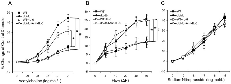
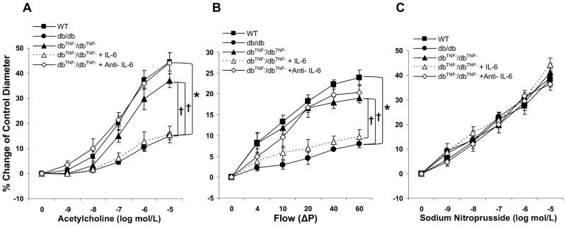
Methods: The studies used wild type (WT), diabetic mice (db/db), db/db null for TNF (dbTNF-/dbTNF-), and db/db mice treated with neutralizing antibody to IL-6 (anti-IL-6). Endothelium-dependent (acetylcholine [ACh], or luminal flow-induced shear stress) and endothelium-independent (sodium nitroprusside [SNP]) vasodilation of isolated and pressurized coronary arterioles were determined. Quantitative PCR, Western blot, and immunofluorescence staining were utilized for mechanistic studies.

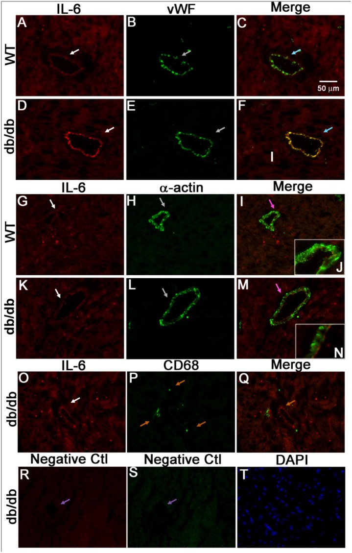
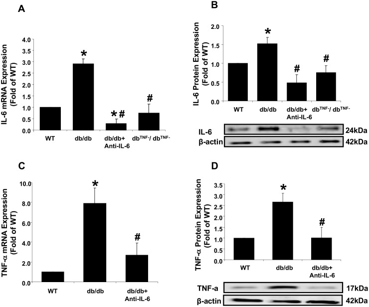
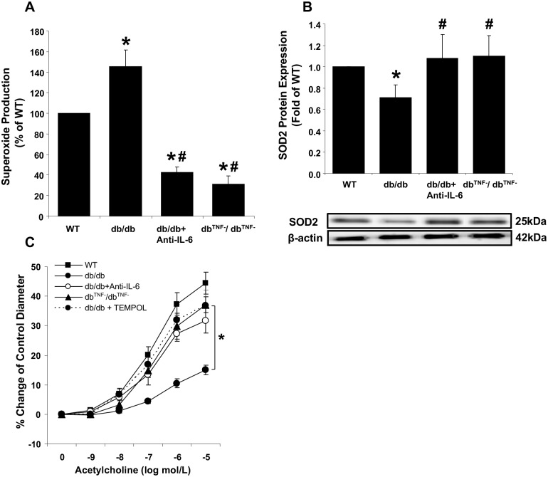
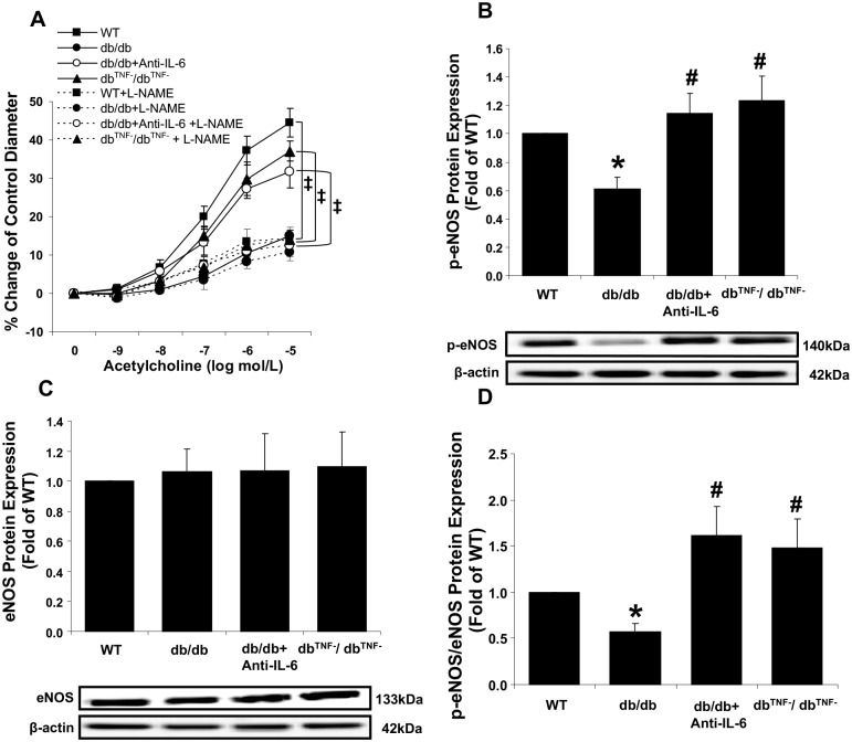
Results: Relative to WT, arteriolar dilation to both ACh and flow was attenuated in db/db mice and dbTNF-/dbTNF-. Treatment of dbTNF-/dbTNF- and db/db mice with anti-IL-6 improved arteriolar dilation compared to db/db mice. Immunofluorescence staining illustrated localization of IL-6 within the endothelial cells of coronary arterioles. In db/db mice, mRNA and protein expression of IL-6 and superoxide (O2-) production were higher, but reduced by anti-IL-6 treatment. Also, in db/db mice, mRNA and protein expression of TNF-α suppressed by the anti-IL-6 treatment and the reduced expression of mRNA and protein expression of IL-6 by the genetic deletion of TNF-α both supported a reciprocal regulation between TNF-α and IL-6. Superoxide dismutase 2 (SOD2) expression and phosphorylation of eNOS (p-eNOS/eNOS) were lower in db/db mice coronary arterioles and were restored in db/db+Anti-IL-6 and dbTNF-/dbTNF- mice.

Conclusion: The interactions between TNF-α and IL-6 exacerbate oxidative stress and reduce phosphorylation of eNOS, thereby contributing to coronary endothelial dysfunction in T2D mice.


Conflict of interest statement

Competing Interests: The authors have declared that no competing interests exist.

Figures

Fig 1
Fig 1. Contribution of IL-6 to endothelial dysfunction in T2D.
(A) Isolated coronary arterioles from the WT (n = 11) and db/db mice (n = 13) dilated in a concentration-dependent manner to ACh. ACh-induced endothelium-dependent vasodilation was significantly reduced in the db/db mice compared to the WT mice. Incubation of IL-6 in coronary arterioles from the WT (n = 7) mice attenuated ACh-induced vasodilation. However, neutralizing antibody to IL-6 (n = 6) restored ACh-induced vasodilation of arterioles from the db/db mice. (B) Flow-induced vasodilation of coronary arterioles was impaired in the db/db mice (n = 11) compared to the WT mice (n = 10), but was enhanced by treatment with a neutralizing antibody to IL-6 (n = 6). Further, ex vivo exposure of WT coronary arterioles to IL-6 decreased flow-mediated vasodilation. (C) Vasodilation to the endothelium-independent vasodilator was not significantly different in any of the 4 groups of mice (n = 6~13). Data are shown as mean ± SEM. * p <.05 vs. db/db; # p <.05 vs. db/db+Anti-IL-6.
Fig 2
Fig 2. Interaction between TNF-α and IL-6 in endothelial dysfunction in T2D.
(A) ACh-induced vasodilation in db/db mice null for TNF (dbTNF-/dbTNF-, n = 6) mice was similar to that in the WT mice and significantly higher than in the db/db mice. IL-6 attenuated ACh-induced vasodilation in dbTNF-/dbTNF- mice (n = 6) to the levels observed in the db/db mice. (B) Flow-induced vasodilation in the db/db mice (n = 13) was impaired while it was preserved in dbTNF-/dbTNF- mice (n = 6) which was similar to the WT mice (n = 11). Exogenous IL-6 decreased flow-mediated vasodilation of dbTNF-/dbTNF- mice coronary arteriole (n = 6). (C) Endothelium-independent vasodilator responses to SNP were similar across all groups (n = 6~13). Data are shown as mean ± SEM. * p <.05 vs. db/db; † p <.05 vs. dbTNF-/dbTNF-.
Fig 3
Fig 3. IL-6 localized to endothelial cells of coronary arterioles.
Dual fluorescence combining IL-6 with markers for endothelial cells [von Willebrand factor (vWF)], vascular smooth muscle cells (α-actin) and macrophages (CD68). (A, B, and C) Dual labeling of IL-6 (red) and vWF (green) in control mouse heart tissues. (D, E, and F) Dual labeling of IL-6 (red) and vWF (green) in db/db mouse heart tissues. Arrows in C and F show the colocalization of IL-6 and endothelial cells (yellow) in the control and db/db mice. (G, H, and I) Dual labeling of IL-6 (red) and α-actin (green) in control mouse heart tissues. (K, L, and M) Dual labeling of IL-6 (red) and α-actin (green) in heart tissues of db/db. Arrows in I and M show the α-actin staining with absence of IL-6 staining in the control and db/db mice. Inserts in I (J) and in M (N) show the higher magnification indicated by arrows in I and M. O, P, and Q, dual labeling of IL-6 (red) and marker of macrophage (green) in db/db mouse heart tissues. Arrows in Q show the specific CD68 staining with absence of IL-6 staining. (R and S) Negative control: arrows show the absence of staining in vessels with control IgG and without primary antibodies. (T) Staining of nuclei with DAPI (blue) in heart tissues of the db/db mice. The immunostaining results suggest that in the hearts of type 2 diabetic mice, IL-6 is mainly localized in endothelial cells, but not in vascular smooth muscle cells and macrophages. Magnification×40. Data shown are representative of 4 separate experiments.
Fig 4
Fig 4. Interactive inhibition between IL-6 and TNF-α expression in diabetic endothelial cells.
(A) mRNA expression of IL-6 was higher (3.6-fold) in the db/db mice compared to the WT mice, and significantly attenuated in the db/db mice treated with anti-IL-6 and in diabetic mice null for TNF-α (dbTNF-/dbTNF-). (B) Protein levels of IL-6 were higher in the db/db mice compared to the WT mice, but lower in the anti-IL-6 antibody-treated and dbTNF-/dbTNF- mice. (C) TNF-α mRNA expression was approximately 8-fold higher in the db/db mice compared to the WT mice, but significantly lower after treatment with the IL-6 neutralizing antibody. (D) TNF-α protein levels were significantly higher in the db/db mice compared to the WT mice, but decreased in the diabetic mice receiving neutralizing antibody to IL-6 (n = 6~11). Data are shown as mean ± SEM. * p <.05 vs. WT; # p <.05 vs. db/db.
Fig 5
Fig 5. Effect of ROS on coronary endothelial dysfunction in T2D.
(A) O2- production was significantly higher in db/db mouse hearts compared to the WT mice, but attenuated in the anti-IL-6 treated db/db or null for TNF-α (dbTNF-/dbTNF-). (n = 6~11) (B) Protein levels of SOD2 were reduced in db/db mouse hearts, but restored to the level of the WT mice in both the anti-IL-6 db/db treated db/db mice and db/db mice null for TNF-α. (n = 6~11) (C) Incubation of TEMPOL restored endothelial function in db/db (n = 8) to the levels observed in dbTNF-/dbTNF- or WT mice. Data are shown as mean ± SEM. * p <.05 vs. WT, # p <.05 vs. db/db.
Fig 6
Fig 6. Role of eNOS in endothelial dysfunction in diabetic mouse coronary arterioles.
(A) Treatment with the eNOS inhibitor, L-NAME, attenuated the improved endothelial function by inhibition of either TNF-α (n = 6) or IL-6 in the db/db mice (n = 10), but not in the db/db mice (n = 8). (B) Levels of p-eNOS protein were significantly decreased in db/db mouse hearts, but were restored in the anti-IL-6 treated db/db mice or db/db mice null for TNF-α null for at levels similar to the WT mice. (C) No differences in total eNOS protein levels were observed between groups (n = 6~11). (D) The ratio of p-eNOS to eNOS protein expression was reduced in db/db mouse hearts compared to the WT mice, while the ratios in the anti-IL-6 treated db/db mice and null for TNF-α were significantly higher than db/db (n = 6~11). Data are shown as mean ± SEM. * p <.05 vs. WT; # p <.05 vs. db/db; p <.05 vs. WT+L-NAME.

References

    1. Pencina MJ, D'Agostino RB Sr., Larson MG, Massaro JM, Vasan RS. Predicting the 30-year risk of cardiovascular disease: the framingham heart study. Circulation. 2009;119(24):3078–84. Epub 2009/06/10. doi: 10.1161/CIRCULATIONAHA.108.816694 . - DOI - PMC - PubMed
    1. Meigs JB. Epidemiology of type 2 diabetes and cardiovascular disease: translation from population to prevention: the Kelly West award lecture 2009. Diabetes care. 2010;33(8):1865–71. Epub 2010/07/30. doi: 10.2337/dc10-0641 . - DOI - PMC - PubMed
    1. Vanhoutte PM. Endothelial dysfunction: the first step toward coronary arteriosclerosis. Circulation journal: official journal of the Japanese Circulation Society. 2009;73(4):595–601. Epub 2009/02/20. . - PubMed
    1. Cai H, Harrison DG. Endothelial dysfunction in cardiovascular diseases: the role of oxidant stress. Circulation research. 2000;87(10):840–4. Epub 2000/11/14. . - PubMed
    1. Gamez-Mendez AM, Vargas-Robles H, Rios A, Escalante B. Oxidative Stress-Dependent Coronary Endothelial Dysfunction in Obese Mice. PloS one. 2015;10(9):e0138609 Epub 2015/09/19. doi: 10.1371/journal.pone.0138609 . - DOI - PMC - PubMed