. 2018 Jan 1;27(1):147-159.
doi: 10.1093/hmg/ddx392.

Changes in extracellular matrix cause RPE cells to make basal deposits and activate the alternative complement pathway

Affiliations

Changes in extracellular matrix cause RPE cells to make basal deposits and activate the alternative complement pathway

Rosario Fernandez-Godino et al. Hum Mol Genet. .

Abstract

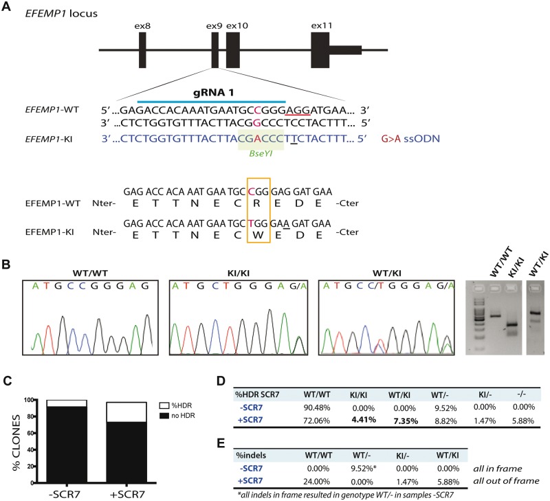
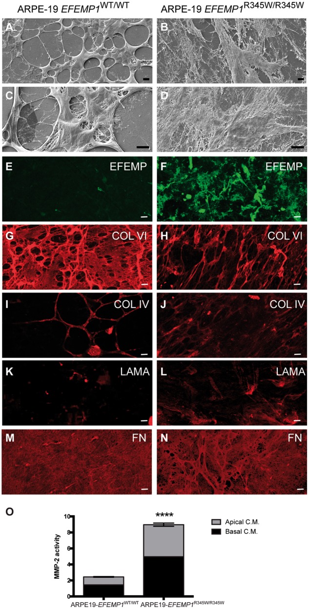
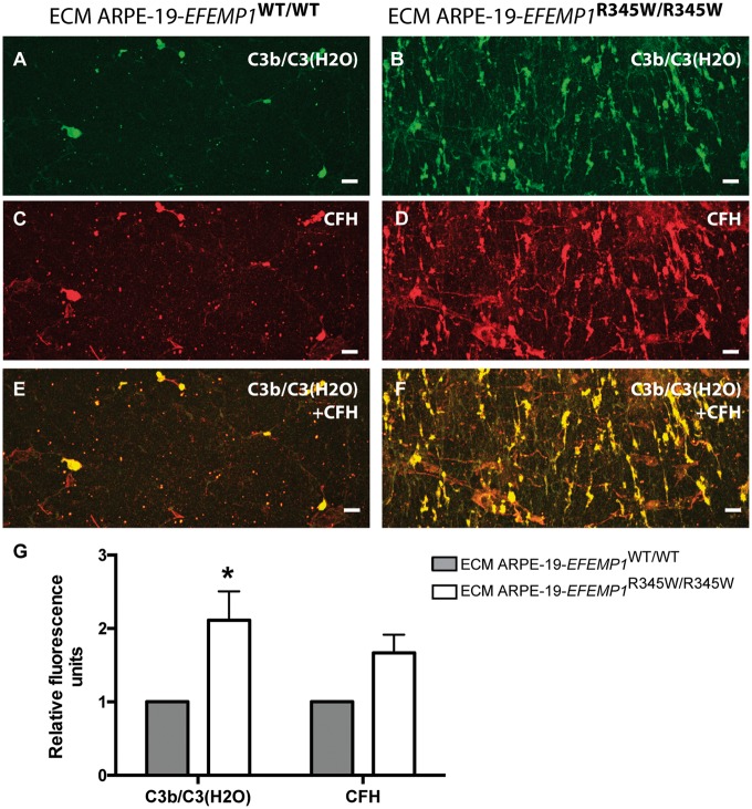
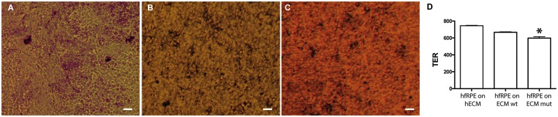
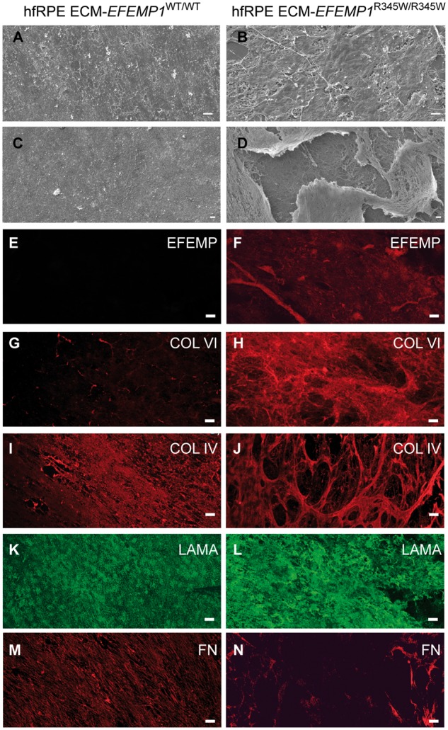
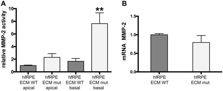
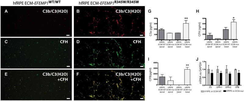
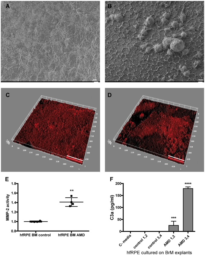
The design of efficient therapies for age-related macular degeneration (AMD) is limited by our understanding of the pathogenesis of basal deposits, which form between retinal pigment epithelium (RPE) and Bruch's membrane (BrM) early in disease, and involve activation of the complement system. To investigate the roles of BrM, RPE and complement in an AMD, we generated abnormal extracellular matrix (ECM) using CRISPR-edited ARPE-19 cells. We introduced to these cells the p.R345W mutation in EFEMP1, which causes early-onset macular degeneration. The abnormal ECM binds active complement C3 and causes the formation of basal deposits by normal human fetal (hf)RPE cells. Human fetal RPE (hfRPE) cells grown on abnormal ECM or BrM explants from AMD donors show chronic activation of the alternative complement pathway by excessive deposition of C3b. This process is exacerbated by impaired ECM turnover via increased matrix metalloproteinase-2 activity. The local cleavage of C3 via convertase-independent mechanisms can be a new therapeutic target for early AMD.


Figures

Figure 1.
Figure 1.
Knock-in the mutation p.R345W in the EFEMP1 gene via CRISPRCas9. (A) Design of guide RNA (gRNA) and single strand oligonucleotide donor (ssODN) to knock-in the mutation c.1033C>T in the exon 9 of the EFEMP1 gene in ARPE-19 cells via CRISPRCas9. PAM sequence underlined in red. Change G>A in the ssODN creates a new restriction site for the BseYI enzyme (highlighted in green). (B) Sanger sequence and agarose gel of successfully edited and isolated clones with genotype WT/WT (wild-type), KI/KI (two copies of the mutation, one per allele) and WT/KI (only one allele has the mutation). (C) Percentage of clones that had the mutation, comparing untreated (−SCR7) and treated (+SCR7) with the DNA ligase IV inhibitor SCR7. (D) Quantification of edited clones according to their genotype: WT/WT: both alleles wild-type; KI/KI: both alleles mutant; WT/KI: one allele mutant and one WT; WT/−: one allele wild-type and one allele with indels that result in null allele; KI/−: one allele with the mutation and one allele with indels that result in null allele; −/−: both alleles have indels that result in null alleles. (E) Percentage of clones treated with or without SCR7 which alleles had indels in frame or out of frame.
Figure 2.
Figure 2.
Edited ARPE-19-EFEMP1R345/R345W cells make abnormal ECM. Decellularized transwells of ARPE-19 wild-type (left panels) and ARPE-19-EFEMP1R345/R345W (right panels) cultures after 4 weeks. SEM images show normal ECM (A and C) versus abnormal ECM (B and D) made by wild-type and mutant cells respectively. Immunostaining with antibodies for EFEMP1 (E and F), Col VI (G and H), Col IV (I and J), laminin (K and L) and fibronectin (M and N) show the abnormal structure of the ECM. Scale bars (A–N): 10 µm. (O) Relative MMP-2 activity measured by zymography in apical and basal conditioned media of ARPE-19-EFEMP1WT/WT and ARPE-19-EFEMP1R345/R345W cells (data represented as mean ±SD; t-test; ****P < 0.0001).
Figure 3.
Figure 3.
Abnormal ECM anchors C3b and CFH. Decellularized transwells of ARPE-19 wild-type (left panels) and ARPE-19-EFEMP1R345/R345W (right panels) cultures. Exposed ECM immunostained with antibodies for (A and B) C3b/C3(H2O) and (C and D) CFH. (E and F) Co-staining for C3b/C3(H2O) and CFH. Scale bars 50 µm. (G) Average quantification of C3b/C3(H2O) and CFH fluorescent labeling comparing exposed ECMs (data represented as mean ± SD. ANOVA, *P < 0.05).
Figure 4.
Figure 4.
HfRPE grown on normal versus abnormal ECM. Brightfield microscopy of hfRPE cells grown for 2 weeks on (A) commercial human ECM, ECM made by (B) ARPE-19 wild-type or (C) ARPE-19-EFEMP1R345/R345W cells. Scale bars 10 µm. (D) TER measured in hfRPE cell cultured on human ECM, ECM made by ARPE-19 wild-type or ARPE-19-EFEMP1R345/R345W cells for 2 weeks (n = 3 cultures/ECM type. t-test, *P < 0.05).
Figure 5.
Figure 5.
HfRPE cells grown on abnormal ECM make basal deposits. (A and C) hfRPE cells grown for 2 weeks on ECM made by ARPE-19 wild-type cells were decellularized and imaged by SEM. Images show the remaining exposed normal ECM made by hfRPE cells. (B and D) hfRPE cells grown for 2 weeks on ECM made by ARPE-19-EFEMP1R345/R345W cells were decellularized and imaged by SEM. Images show the remaining thick basal deposits made by hfRPE cells. In some of these areas, the outer layer of the deposits is lifted and a network of crosslinking ECM fibers can be observed underneath. Immunostaining of the exposed ECM and deposits with antibodies for EFEMP1 (E and F), Col VI (G and H), Col IV (I and J), laminin (K and L) and fibronectin (M and N). Immunolabeling of deposits revealed a very strong signal for collagen VI, and the distribution of both collagen VI and IV was heterogeneous and disposed as a tridimensional network of crosslinked bundles of collagen fibers. The data show that the composition of the deposits made by hfRPE grown on abnormal ECM is similar to the composition basal deposits in AMD patients. Scale bars A–D: 10 µm, E–N: 50 µm.
Figure 6.
Figure 6.
Abnormal ECM alters the ECM turnover. (A) MMP-2 activity measured by zymography in apical and basal conditioned media of hfRPE cells cultured on normal (wt) or abnormal (mut) ECM made by ARPE-19-EFEMP1R345/R345W. (B) mRNA levels of MMP-2 measured in the same cultures, normalized to GAPDH (data represented as mean ± SEM; t-test, **P < 0.01; n = 4/ECM type).
Figure 7.
Figure 7.
Abnormal ECM causes complement activation by normal hfRPE cells. hfRPE cultures grown on ECM made by ARPE-19 wild-type or ARPE-19-EFEMP1R345/R345W were decellularized and immunostained with antibodies for (A and B) C3b/C3(H2O), (C and D) CFH or (E and F) both C3b/C3(H2O) and CFH. Scale bars: 50µm. (G) C3a, (H) CFH and (I) CFB measured by ELISA in apical and basal conditioned media of hfRPE cells grown on ECM made by ARPE-19 wild-type (ECM WT) or ARPE-19-EFEMP1R345/R345W (ECM mut) in the absence of serum (data represented as mean ± SD. ANOVA, n = 4 per type, *P < 0.005, **P < 0.001). (J) mRNA levels of complement components from the alternative pathway measured in hfRPE cells grown on normal (gray) or abnormal (black) ECM made by wild-type or mutant ARPE-19 cells, respectively.
Figure 8.
Figure 8.
BrM with AMD alters ECM turnover and causes complement activation by normal hfRPE cells. SEM of BrM from (A) normal and (B) AMD donors. Thick ECM network and drusen-like deposits are visible in the BrM of AMD donors. Representative 3D reconstruction of BrM explants from (C) normal and (D) AMD donors immunostained for collagen IV, and imaged by confocal microscope. Quantification of (C) MMP-2 activity and (D) C3a in conditioned media from hfRPE cells cultured on BrM–choroid–sclera explants from donors without AMD (control 1–4) and with AMD (AMD 1–4). C-media: no cells (data represented as mean ± SD. n = 4 explants/type. ANOVA, ****P < 0.0001, ***P < 0.001, **P < 0.01). Scale bars: (A, B)=10 µm, (C, D)=50 µm.

References

    1. Miller J.W. (2013) Age-related macular degeneration revisited–piecing the puzzle: the LXIX Edward Jackson memorial lecture. Am. J. Ophthalmol., 155, 1–35.e13. - PubMed
    1. Evans J.R., Lawrenson J.G. (2012) Antioxidant vitamin and mineral supplements for slowing the progression of age-related macular degeneration. Cochrane Database Syst. Rev., 11, CD000254. - PubMed
    1. Newsome D.A., Huh W., Green W.R. (1987) Bruch's membrane age-related changes vary by region. Curr. Eye Res., 6, 1211–1221. - PubMed
    1. Nita M., Strzalka-Mrozik B., Grzybowski A., Mazurek U., Romaniuk W. (2014) Age-related macular degeneration and changes in the extracellular matrix. Med. Sci. Monit., 20, 1003–1016. - PMC - PubMed
    1. Curcio C.A., Johnson M. (2013) Structure, function, and pathology of Bruch's membrane. Retina, 1, 465–481.

Publication types