Review
. 2017 Nov 1;96(3):651-666.
doi: 10.1016/j.neuron.2017.09.055.

Mitostasis in Neurons: Maintaining Mitochondria in an Extended Cellular Architecture

Affiliations
Review

Mitostasis in Neurons: Maintaining Mitochondria in an Extended Cellular Architecture

Thomas Misgeld et al. Neuron. .

Abstract

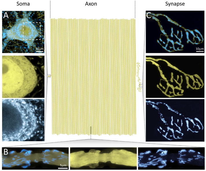
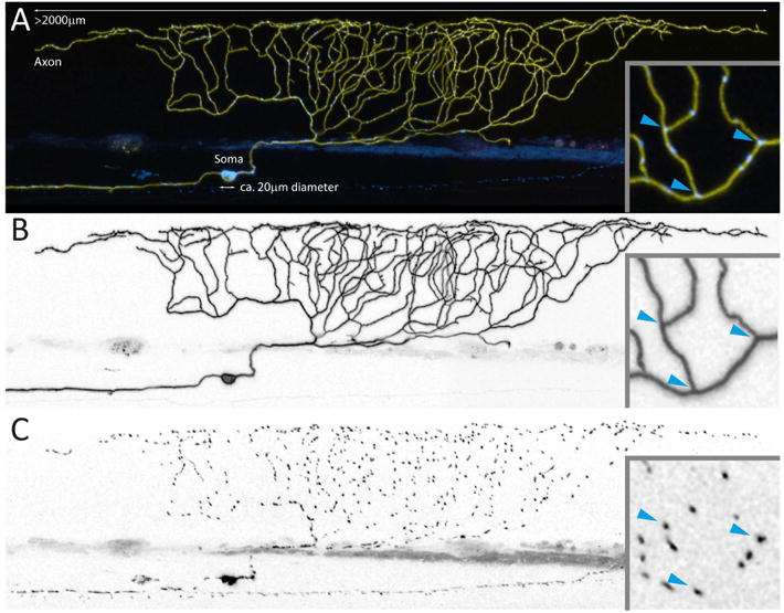
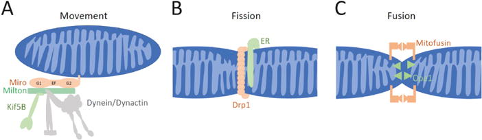
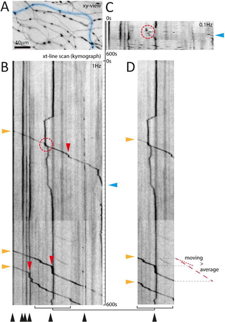
Neurons have more extended and complex shapes than other cells and consequently face a greater challenge in distributing and maintaining mitochondria throughout their arbors. Neurons can last a lifetime, but proteins turn over rapidly. Mitochondria, therefore, need constant rejuvenation no matter how far they are from the soma. Axonal transport of mitochondria and mitochondrial fission and fusion contribute to this rejuvenation, but local protein synthesis is also likely. Maintenance of a healthy mitochondrial population also requires the clearance of damaged proteins and organelles. This involves degradation of individual proteins, sequestration in mitochondria-derived vesicles, organelle degradation by mitophagy and macroautophagy, and in some cases transfer to glial cells. Both long-range transport and local processing are thus at work in achieving neuronal mitostasis-the maintenance of an appropriately distributed pool of healthy mitochondria for the duration of a neuron's life. Accordingly, defects in the processes that support mitostasis are significant contributors to neurodegenerative disorders.

Keywords: axonal transport; homeostasis; mitochondria; mitophagy; neurons.


Figures

Figure 1
Figure 1. Neuronal Mitochondria in a Mouse Neuron
The extended geometry of neurons poses special challenges for maintaining mitostasis – here illustrated by a schematic attempt to draw a mammalian motor neuron to scale, with the soma to the left, the axon in the center, and the synapse-studded branches to the right (modified from (Devor, 1999)). A small soma (A) with a limited pool of mitochondria has to supply a vast axonal arbor that contains a far larger population of unevenly distributed mitochondria. At the same time, neuronal compartments with specific bioenergetic needs and hence accumulations of neuronal mitochondria, such as paranodes (B) or presynaptic terminals (C), need to be served.
Figure 2
Figure 2. Neuronal Mitochondria in a Zebrafish Neuron
Smaller model organisms, such as flies, nematodes and zebrafish larvae (shown here), allow studying mitostatic mechanisms in a slightly less complex in vivo environment than in mammals. (A) depicts a zebrafish sensory Rohon-Beard neuron (96 hours post fertilization), which expresses a yellow fluorescent protein in the membrane and a cyan fluorescent protein in the mitochondrial matrix – giving an impression of the relative size of soma and axon (Plucinska et al., 2012). Optical accessibility allows entire axonal arbors (B) to be visualized and interrogated for mitochondrial distribution (C) and turnover. Even in these unmyelinated axons lacking synapses, mitochondria do not appear to be randomly distributed – for instance, many mitochondria reside at branch points (blue arrowheads in inset).
Figure 3
Figure 3. Mechanisms for Mitochondrial Dynamics in Neurons
(A) To move along microtubules in both the anterograde direction (towards the + ends of microtubules) and retrograde (towards – ends), mitochondria employ a motor/adaptor complex that links Kif5b and the dynein/dynactin complex to the mitochondrial surface (Saxton and Hollenbeck, 2012; Schwarz, 2013). (B) Fission of both the inner and outer mitochondrial membranes is mediated by a ring of the dynamin-like GTPase Drp1. In many cases, fission appears to be initiated by a contact with the endoplasmic reticulum (ER). (C) Full fusion of mitochondria has two components. For the outer membranes to fuse, Mitofusins must be present on the membranes of both mitochondria and their interaction initiates the fusion. The inner membranes undergo a separate fusion that is mediated by Opa1.
Figure 4
Figure 4. Challenges in Measuring Mitochondrial Movement
The parameters of motility are often difficult to compare, as different studies may choose to quantify the flux through a point, the percent of mitochondria that move, or the fraction of time that mitochondria spend in motion. Moreover, parameters that would appear to be fundamental properties are in fact operationally defined. (A–D) illustrate how the imaging protocol can affect analysis of kymographs from sensory axons in the skin of larval zebrafish (Plucinska et al., 2012). (A) xy-view of the region of interest imaged at 1 Hz. (B) A fully resolved kymograph along the blue line in A reveals different mitochondrial populations – stationary as vertical lines (black arrowheads) vs. moving as angled lines (three anterograde mitochondria; orange arrowheads), with short vertical segments (i.e. pauses; red arrowheads). Such pauses explain the difference between average speed (dashed red line next to D) vs. ‘moving’ speed (excluding pauses; dotted red line). (C) Temporal under-sampling (0.1 Hz was mimicked), leads to broken tracks that are difficult to follow (red circle) and obscures small pauses (compare track with cyan triangle). (D) The size of the field of view can affect the measured motile fraction (B, 3/9 mitochondria = 33% vs. D, 3/4 = 75%), as can the recording length because the stationary count will stay constant as moving mitochondria continue to pass through the field (not shown; consider the motile fraction in the upper, or lower half of B: 11% and 25%, respectively). Together these sampling discrepancies, but also signal-to-noise and movement artifacts, explain some of the reported ranges of transport parameters (Table 1). Not illustrated here are the effects of axonal branching or of fission and fusion, which further complicate such measurements.
Figure 5
Figure 5. Mechanisms of Mitochondrial Protein Turnover
The mitochondrial quality is preserved by four mechanisms illustrated in order of their increasing scale. (A) Individual misfolded or damaged proteins are degraded by any of several mitochondrial proteases. (B) Mitochondrial components can be shed from the organelle in MDVs that bud from the organelle and can contain either only outer membrane components or also components of the inner membrane and matrix. (C) Mitophagy can engulf mitochondria, whose membrane potential or protein misfolding have triggered the PINK1/Parkin pathway to add phospho-ubiquitin to the outer membrane and thereby induce formation of autophagosomal membranes. The autophagosome will subsequently fuse with a lysosome. (D) Macroautophagy occurs in growth cones and can sweep up mitochondria along with other cytoplasmic components. Once formed, the autophagosome moves retrograde and fuses with lysosomes in the axon.

References

    1. Ahmed AU, Fisher PR. Import of nuclear-encoded mitochondrial proteins: a cotranslational perspective. Int Rev Cell Mol Biol. 2009;273:49–68. - PubMed
    1. Alexander C, Votruba M, Pesch UE, Thiselton DL, Mayer S, Moore A, Rodriguez M, Kellner U, Leo-Kottler B, Auburger G, et al. OPA1, encoding a dynamin-related GTPase, is mutated in autosomal dominant optic atrophy linked to chromosome 3q28. Nat Genet. 2000;26:211–215. - PubMed
    1. Alston CL, Rocha MC, Lax NZ, Turnbull DM, Taylor RW. The genetics and pathology of mitochondrial disease. Journal of Pathology. 2017;241:236–250. - PMC - PubMed
    1. Amiri M, Hollenbeck PJ. Mitochondrial biogenesis in the axons of vertebrate peripheral neurons. Dev Neurobiol. 2008;68:1348–1361. - PMC - PubMed
    1. Andreyev AY, Kushnareva YE, Murphy AN, Starkov AA. Mitochondrial ROS Metabolism: 10 Years Later. Biochemistry (Mosc) 2015;80:517–531. - PMC - PubMed