Review
. 2017 Nov 1;96(3):680-696.
doi: 10.1016/j.neuron.2017.10.006.

Transcellular Nanoalignment of Synaptic Function

Affiliations
Review

Transcellular Nanoalignment of Synaptic Function

Thomas Biederer et al. Neuron. .

Abstract

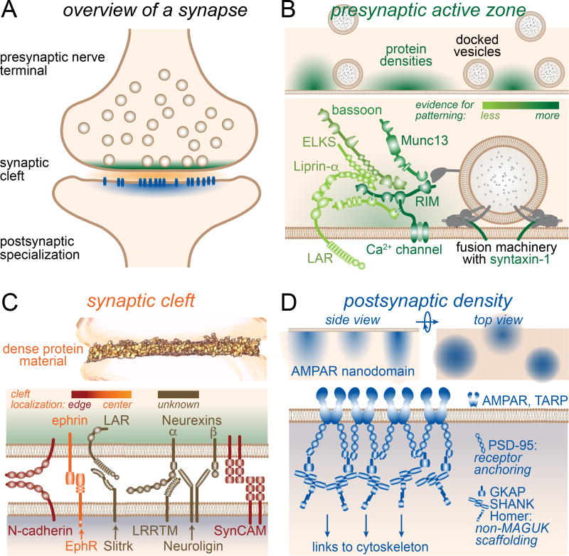
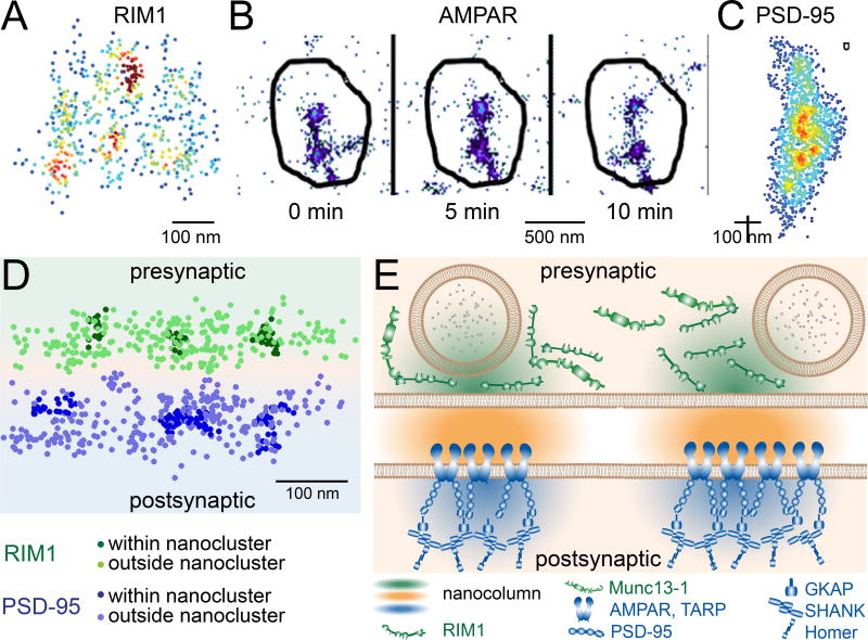
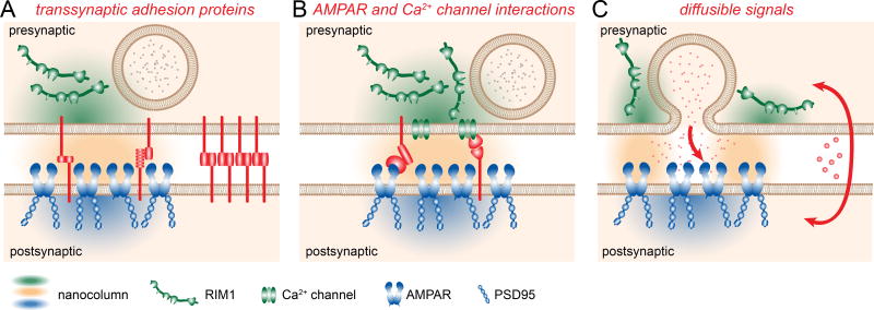
At each of the brain's vast number of synapses, the presynaptic nerve terminal, synaptic cleft, and postsynaptic specialization form a transcellular unit to enable efficient transmission of information between neurons. While we know much about the molecular machinery within each compartment, we are only beginning to understand how these compartments are structurally registered and functionally integrated with one another. This review will describe the organization of each compartment and then discuss their alignment across pre- and postsynaptic cells at a nanometer scale. We propose that this architecture may allow for precise synaptic information exchange and may be modulated to contribute to the remarkable plasticity of brain function.

Keywords: active zone; nanocolumn; postsynaptic density; synapse; synaptic cleft.


Figures

Figure 1
Figure 1. The machines of the synapse
(A) Overview of an excitatory synapse with the active zone material (green), the synaptic cleft material (orange) and the postsynaptic density (blue) highlighted in a color code that is maintained throughout the manuscript. (B) Patterning (top) and molecular components (bottom, adapted from Kaeser et al., 2011) of the presynaptic active zone area. (C) 3D tomogram of the protein complexes in side view (top, adapted from Perez de Arce et al., 2015) and its molecular components (bottom) of the synaptic cleft. Individual cleft proteins are discussed in the text. Notably, proteinaceous material within the cleft is distinctly distributed, with the highest density in the outer ring. (D) Lateral patterning of the postsynaptic density in side and top view (top) and patterned molecular components of a single cluster (bottom) of the postsynaptic density. The PSD material including receptors is organized in ∼2–3 distinct clusters per synapse and is also layered vertically into functionally relevant zones.
Figure 2
Figure 2. Evidence for alignment
(A) Clustered distribution of RIM1 within a single active zone, visualized via STORM and rotated to view along the synaptic axis parallel to the presynaptic plasma membrane. Points indicate position of localized molecules, and color encodes the relative number of other nearby molecules with red being the highest and blue the lowest. (Adapted from Tang et al., 2017.) (B) Clustering of AMPARs visualized by time-lapsed single-molecule mapping. Receptors (points) concentrate in small subdomains of a single PSD (outline). Most receptors are in motion, but the nanoclusters remain stable for long periods. (Adapted from Nair et al., 2013.) (C) Clustered distribution of PSD-95 within a single PSD, displayed as in A. (Adapted from MacGillavry et al., 2013). (D) Trans-synaptic alignment of nanoclustered RIM1 and PSD-95 visualized by 3D dSTORM. The synapse has been rotated to view perpendicular to the synaptic cleft. (E) The “nanocolumn” of protein alignment spanning the two connected neurons. Concentrated RIM helps establish preferential release sites in the active zone which align within 10s of nanometers to clusters of AMPARs. The density of cleft protein (orange) within the nanocolumn has not been measured, but Munc13 co-enriches with RIM in the active zone, and Shank, GKAP, and Homer co-enrich with PSD-95.
Figure 3
Figure 3. Alternative mechanisms of alignment
Three possible mechanisms for trans-synaptic alignment into nanocolumns are direct interactions between trans-synaptic adhesion proteins (A), interactions of cleft proteins with presynaptic Ca2+ channels or postsynaptic receptors (B), or diffusible signals for antero- or retrograde signaling (C). Each group of mechanisms is discussed in the text, and they may account on their own or in any combination for the nanocolumn structure.

References

    1. Acuna C, Liu X, Gonzalez A, Südhof TC. RIM-BPs Mediate Tight Coupling of Action Potentials to Ca<sup>2+</sup>-Triggered Neurotransmitter Release. Neuron. 2015;87:1234–1247. - PubMed
    1. Adesnik H, Nicoll RA, England PM. Photoinactivation of Native AMPA Receptors Reveals Their Real-Time Trafficking. Neuron. 2005;48:977–985. - PubMed
    1. Ahmad-Annuar A, Ciani L, Simeonidis I, Herreros J, Fredj NB, Rosso SB, Hall A, Brickley S, Salinas PC. Signaling across the synapse: a role for Wnt and Dishevelled in presynaptic assembly and neurotransmitter release. J Cell Biol. 2006;174:127–139. - PMC - PubMed
    1. Anderson GR, Aoto J, Tabuchi K, Foldy C, Covy J, Yee AX, Wu D, Lee SJ, Chen L, Malenka RC, Sudhof TC. beta-Neurexins Control Neural Circuits by Regulating Synaptic Endocannabinoid Signaling. Cell. 2015;162:593–606. - PMC - PubMed
    1. Andrews-Zwilling YS, Kawabe H, Reim K, Varoqueaux F, Brose N. Binding to Rab3A-interacting molecule RIM regulates the presynaptic recruitment of Munc13-1 and ubMunc13-2. The Journal of biological chemistry. 2006;281:19720–19731. - PubMed

MeSH terms