. 2017 Nov 2;17(1):709.
doi: 10.1186/s12885-017-3709-3.

Therapy response testing of breast cancer in a 3D high-throughput perfused microfluidic platform

Affiliations

Therapy response testing of breast cancer in a 3D high-throughput perfused microfluidic platform

Henriette L Lanz et al. BMC Cancer. .

Abstract

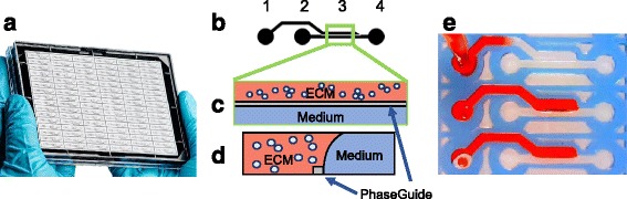
Background: Breast cancer is the most common invasive cancer among women. Currently, there are only a few models used for therapy selection, and they are often poor predictors of therapeutic response or take months to set up and assay. In this report, we introduce a microfluidic OrganoPlate® platform for extracellular matrix (ECM) embedded tumor culture under perfusion as an initial study designed to investigate the feasibility of adapting this technology for therapy selection.

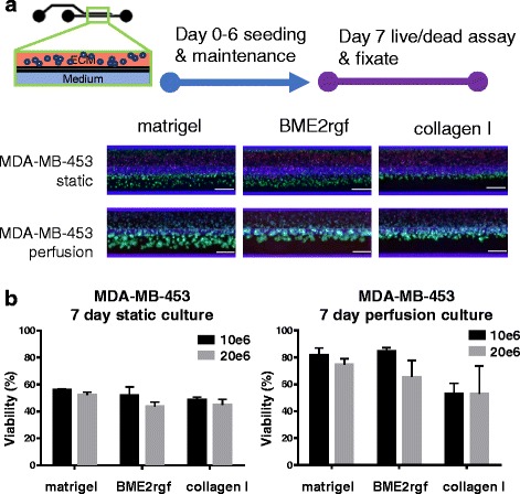
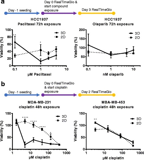
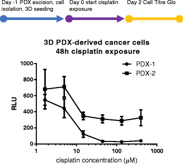
Methods: The triple negative breast cancer cell lines MDA-MB-453, MDA-MB-231 and HCC1937 were selected based on their different BRCA1 and P53 status, and were seeded in the platform. We evaluate seeding densities, ECM composition (Matrigel®, BME2rgf, collagen I) and biomechanical (perfusion vs static) conditions. We then exposed the cells to a series of anti-cancer drugs (paclitaxel, olaparib, cisplatin) and compared their responses to those in 2D cultures. Finally, we generated cisplatin dose responses in 3D cultures of breast cancer cells derived from 2 PDX models.

Results: The microfluidic platform allows the simultaneous culture of 96 perfused micro tissues, using limited amounts of material, enabling drug screening of patient-derived material. 3D cell culture viability is improved by constant perfusion of the medium. Furthermore, the drug response of these triple negative breast cancer cells was attenuated by culture in 3D and differed from that observed in 2D substrates.

Conclusions: We have investigated the use of a high-throughput organ-on-a-chip platform to select therapies. Our results have raised the possibility to use this technology in personalized medicine to support selection of appropriate drugs and to predict response to therapy in a real time fashion.

Keywords: Organ-on-a-chip; P53 and BRCA1; Personalized medicine; Triple negative.


Conflict of interest statement

Ethics approval and consent to participate

The generation of the breast cancer PDX models were described previously [37]. The Mayo Clinic Institutional Animal Care and Use Committee (IACUC) reviewed and approved all of the mouse experiments for the PDX tumors used in this study.

Consent for publication

Not applicable

Competing interests

Paul Vulto, Jos Joore, Thomas Hankemeier and Sebastiaan J Trietsch have ownership interest in Mimetas B.V, which has developed the technology reported in this publication. Henriette Lanz, Anthony Saleh, Bart Kramer and Chee Ping Ng are employees of Mimetas B.V.

Publisher’s Note

Springer Nature remains neutral with regard to jurisdictional claims in published maps and institutional affiliations.

Figures

Fig. 1
Fig. 1
Microtiter cancer-on-a-chip plate for 3D breast cancer therapy response testing. a Photo of OrganoPlate® platform consisting of 96 perfusable microfluidic chambers in parallel. b Closeup, (c) top and (d) side view of an individual chamber consisting of an ECM channel and a Medium channel. Cells are premixed into a gel solution, loaded into the ECM channel by capillary action and allowed to polymerize before the introduction of medium into the adjacent Medium channel for culture. PhaseGuide™ allows the gel solution to be pinned during the loading and polymerization step, thereby allowing support-free and unhindered exchange with the medium. e Photo demonstrates the filling of the ECM channel using a red dye
Fig. 2
Fig. 2
Culture optimization in the microtiter microfluidic platform. Up to 96 multiple conditions such as seeding density, ECM composition, cell types and perfusion can be investigated concurrently. Breast cancer cell line MDA-MB-453 was seeded in three different ECM compositions at two different densities and maintained for 6 days before assessment with a live/dead assay (Calcein AM - green/NucBlue® (Hoechst) - blue/NucRed® (propidium iodide) - Red). Scale bar = 400 μm. a Epifluorescence microscopy images showing different morphologies and viabilities of MDA-MB-453 in Matrigel®, BME2rgf and collagen I under static and perfusion conditions at a seeding density of 10*106 cells/mL. b Graphs quantifying the effect of ECM (Matrigel® vs BME2rgf vs collagen I), seeding density (10*106 cells/mL, black, vs 20*106 cells/mL, grey), and static vs perfusion culture on the viability (represented as % of total cells) of MDA-MB-453 cells. Total cell number was determined by nuclear count (Hoechst staining). Total number of dead cells was determined by positive propidium iodide staining. Viable cells was set at total cell number minus dead cell count
Fig. 3
Fig. 3
Screening studies of breast cancer cell lines in microfluidic culture. a For paclitaxel and olaparib studies, HCC-1937 were seeded 3D in the OrganoPlate® and 2D on tissue culture grade plastics 96-well flat bottom plates, cultured for 1 day before 72 h exposure with compounds at the specified concentrations. Viability (as % of total cells) was quantified using an optimized RealTime-Glo™ cell viability assay. (B) MDA-MB-231 and MDA-MB-453 were seeded for 24 h similarly and exposed to cisplatin at various concentrations for 48 h. Symbols: * P ≤ 0.05, ** P ≤ 0.01, *** P ≤ 0.001, **** P ≤ 0.0001 using Tukey’s multiple comparison test (See supplementary documents for p-values and more detailed analysis)
Fig. 4
Fig. 4
Cisplatin exposure of PDX-derived human breast cancer cells in 3D microfluidic culture. Human cancer cells from two different breast cancer PDX avatars were isolated and seeded in 3D in the OrganoPlate® 1 day prior to 48 h cisplatin exposure. Culture viability was quantified using the luminescent CellTiter-Glo® cell viability assay. IC50 were determined based on nonlinear fit of the dose response range as 8,1 μM and 14,8 μM for PDX-1 and PDX-2 respectively
Fig. 5
Fig. 5
Outlook: Work flow for Patient derived xenograft (PDX) vs cancer-on-a-chip drug screening. Compared to PDX drug screening, the compact OrganoPlate® platform is expected to reduce assay time and space, and increase the throughput of screened compounds, leading to improvements in cancer treatment planning and personalized medicine for individual patients

References

    1. Siegel R, Miller K, Jemal A. Cancer statistics, 2015. CA Cancer J Clin [Internet] 2015;65:5–29. doi: 10.3322/caac.21254. - DOI - PubMed
    1. Siegel R, Miller K, Jemal A. Cancer statistics, 2016. CA Cancer J Clin. 2016;66:7–30. doi: 10.3322/caac.21332. - DOI - PubMed
    1. Torre LA, Bray F, Siegel RL, Ferlay J, Lortet-tieulent J, Jemal A. Global cancer statistics, 2012. CA a cancer J Clin [Internet] 2015;65:87–108. doi: 10.3322/caac.21262. - DOI - PubMed
    1. Boyle P. Triple-negative breast cancer: epidemiological considerations and recommendations. Ann Oncol [Internet] 2012;23:8–13. - PubMed
    1. Hurvitz S, Mead M. Triple-negative breast cancer. Curr Opin Obs Gynecol [Internet] 2016;28:59–69. - PubMed

MeSH terms