Comparative Study
. 2018 Jan;38(1):143-153.
doi: 10.1161/ATVBAHA.117.310035. Epub 2017 Nov 2.

Sex Chromosome Complement Defines Diffuse Versus Focal Angiotensin II-Induced Aortic Pathology

Affiliations
Comparative Study

Sex Chromosome Complement Defines Diffuse Versus Focal Angiotensin II-Induced Aortic Pathology

Yasir Alsiraj et al. Arterioscler Thromb Vasc Biol. 2018 Jan.

Abstract

Objective: Aortic pathologies exhibit sexual dimorphism, with aneurysms in both the thoracic and abdominal aorta (ie, abdominal aortic aneurysm [AAA]) exhibiting higher male prevalence. Women have lower prevalence of aneurysms, but when they occur, aneurysms progress rapidly. To define mechanisms for these sex differences, we determined the role of sex chromosome complement and testosterone on the location and progression of angiotensin II (AngII)-induced aortic pathologies.

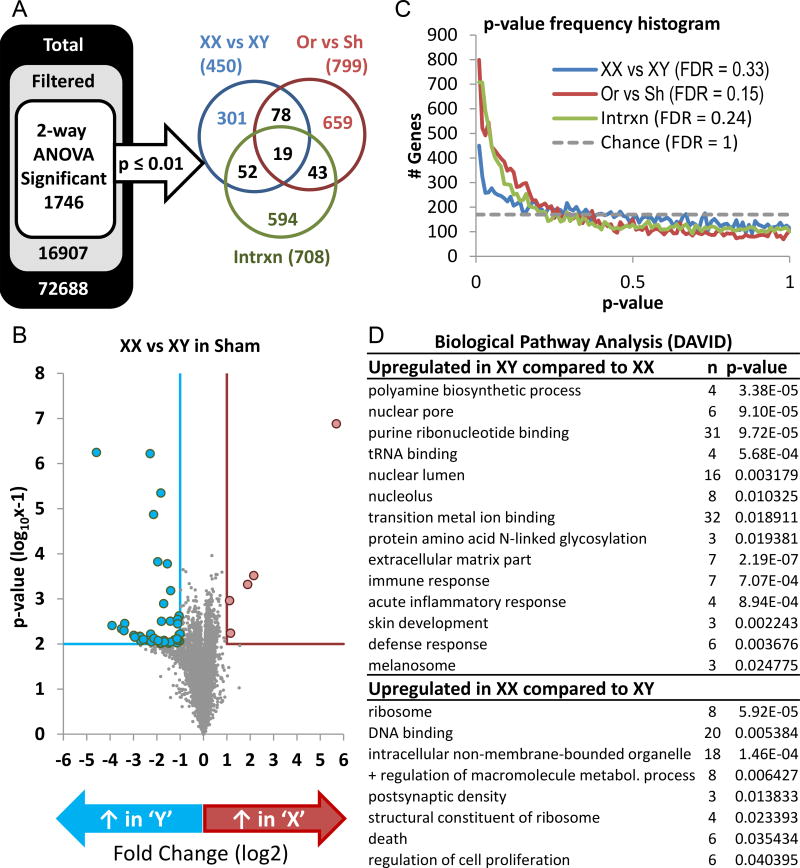
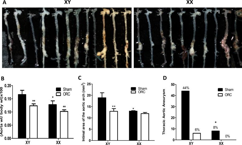
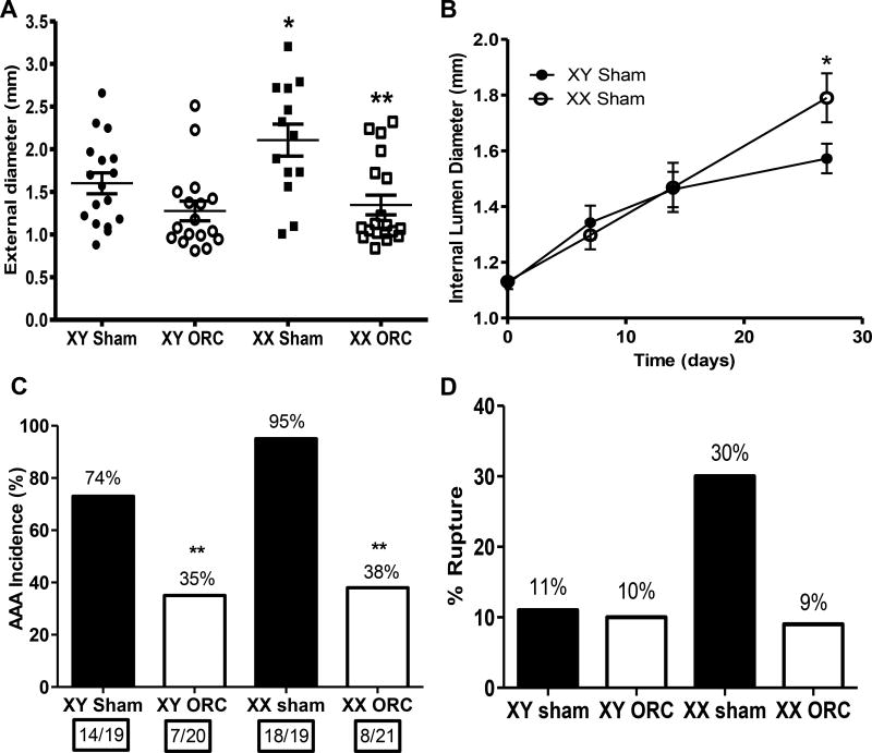
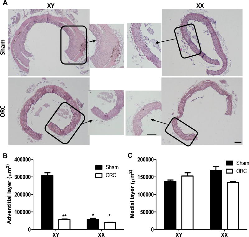
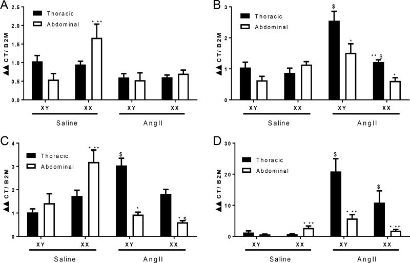
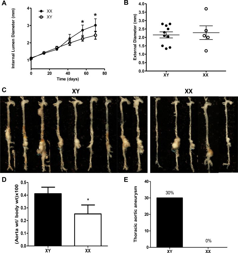
Approach and results: We used transgenic male mice expressing Sry (sex-determining region Y) on an autosome to create Ldlr (low-density lipoprotein receptor)-deficient male mice with an XY or XX sex chromosome complement. Transcriptional profiling was performed on abdominal aortas from XY or XX males, demonstrating 1746 genes influenced by sex chromosomes or sex hormones. Males (XY or XX) were either sham-operated or orchiectomized before AngII infusions. Diffuse aortic aneurysm pathology developed in XY AngII-infused males, whereas XX males developed focal AAAs. Castration reduced all AngII-induced aortic pathologies in XY and XX males. Thoracic aortas from AngII-infused XY males exhibited adventitial thickening that was not present in XX males. We infused male XY and XX mice with either saline or AngII and quantified mRNA abundance of key genes in both thoracic and abdominal aortas. Regional differences in mRNA abundance existed before AngII infusions, which were differentially influenced by AngII between genotypes. Prolonged AngII infusions resulted in aortic wall thickening of AAAs from XY males, whereas XX males had dilated focal AAAs.

Conclusions: An XY sex chromosome complement mediates diffuse aortic pathology, whereas an XX sex chromosome complement contributes to focal AngII-induced AAAs.

Keywords: aneurysm; aorta; pathology; sex chromosomes; testosterone.


Figures

Figure 1
Figure 1
Sex chromosome complement, sex hormones, and an interaction between these factors influence gene expression patterns in abdominal aortas of XY and XX males. A, Total number of probe sets on arrays filtered to retain transcripts with reliable signal intensity (FDR). B, Volcano plot illustrating fold change in gene abundance (x-axis) and statistical significance (y-axis). Genes labelled in blue exhibited significant increase in XY compared to XX abdominal aortas; genes labeled in red exhibited significant increase in XX aortas. C, Frequency histogram of the number of genes (y-axis) found at different p-values (x-axis). Chance is illustrated by the dashed line. D, Biological pathway analysis (DAVID) of gene expression comparing pathways upregulated in XY or XX abdominal aortas. Data are mean ± SEM from n = 4–5 mice/genotype.
Figure 2
Figure 2
An XY sex chromosome complement mediates diffuse aortic vascular disease, while an XX sex chromosome complement is associated with discrete aneurysmal disease in abdominal aortas of male AngII-infused mice. A, Aortas from XY and XX sham-operated males infused with AngII. B, Aorta weight normalized to body weight. C, Area of the aortic arch. D, Incidence of thoracic aortic aneurysms (%) in mice of each genotype and group. Data are mean ± SEM from n = 10–13 mice/genotype/group who survived the 28 day protocol. *, P<0.05 compared to XY within sham-operated determined by two-way ANOVA with sham and genotype as between group factors with Holm-Sidak post hoc analysis (B,C) or by Fisher’s exact test (D). **, P<0.05 compared to sham-operated within genotype determined by two-way ANOVA with sham and genotype as between group factors with Holm-Sidak post hoc analysis.
Figure 3
Figure 3
An XX sex chromosome complement results in focal AAA pathology of increased size compared to XY males infused with AngII. A, Maximal AAA external diameters. Symbols represent individual mice with lines representing mean ± SEM. B, Internal diameters of abdominal aortas at selected intervals during infusion of AngII. Data are mean ± SEM of mice surviving the 28 day infusions. C, AAA incidence (percent above each bar). Numbers in boxes under each bar are mice with an AAA/total number of mice per group. D, % Aneurysm rupture (percent above each bar). *, P<0.05 compared to XY within sham-operated determined by two-way ANOVA with Holm-Sidak post hoc analysis (A,B) or by Fisher’s exact test (C,D). **, P<0.05 compared to sham-operated within genotype determined by two-way ANOVA with Holm-Sidak post hoc analysis (A,B) or by Fisher’s exact test (C,D).
Figure 4
Figure 4
Thoracic aortas from XY males, but not XX males, exhibit adventitial thickening in response to AngII infusions. A, Representative thoracic aorta tissue sections from XY and XX sham-operated 51 and orchiectomized (ORC) males infused with AngII for 28 days. Boxes are areas illustrated at higher magnification. B, Quantification of the adventitial layer of thoracic aorta tissue sections from mice of each group. C, Quantification of the medial layer of thoracic aorta tissue sections from mice of each group. Data are mean ± from 6 tissue sections from n = 3 mice/group. *, P<0.05 compared to XY within treatment group determined by two-way ANOVA with surgery and genotype as between group factors, with Holm-Sidak post hoc analysis. **, P<0.05 compared to sham within genotype determined by two-way ANOVA with surgery and genotype as between group factors, with Holm-Sidak post hoc analysis. Scale bar represents 100 µm.
Figure 5
Figure 5
mRNA abundance of key genes implicated in aneurysm development in thoracic versus abdominal aortas from XY and XX male mice infused with either saline or AngII-infused (1 day). A, Angiotensin converting enzyme (ACE) mRNA abundance. B, Matrix metalloproteinase 2 mRNA abundance. C, Collagen 1a1 mRNA abundance. D, Thrombospondin 1 mRNA abundance. Data are mean ± SEM from n = 5 mice/group/sex. *, P<0.05 compared to thoracic within genotype determined by three-way ANOVA with region, treatment or genotype as between group factors and Holm-Sidak post hoc analysis. **, P<0.05 compared to XY within aortic region determined by three-way ANOVA with region, treatment or genotype as between group factors and Holm-Sidak post hoc analysis. $, compared to saline within region and genotype determined by three-way ANOVA with region, treatment or genotype as between group factors and Holm-Sidak post hoc analysis.
Figure 6
Figure 6
XX males exhibit dilated AAAs while XY males exhibit diffuse aortic disease with prolonged AngII infusions (3 months). A, Internal lumen diameters of suprarenal aorta at selected intervals of AngII infusions. B, Maximal AAA external diameters at study endpoint. Symbols represent individual mice with lines mean ± SEM. C, Aortas from mice in each group at study endpoint. D, Aorta weight normalized to body weight. E, Incidence of thoracic aortic aneurysms in mice of each genotype. Data are mean ± SEM from n = 5–10 mice/group. *, P<0.05 compared to XY determined by Student t-test (A-D) or Fisher’s exact test (E).

Comment in

References

    1. Roman MJ, Devereux RB, Preiss LR, et al. Gen TACI. Associations of age and sex with marfan phenotype: The national heart, lung, and blood institute gentac (genetically triggered thoracic aortic aneurysms and cardiovascular conditions) registry. Circulation. Cardiovascular genetics. 2017;10 - PMC - PubMed
    1. Cheung K, Boodhwani M, Chan KL, Beauchesne L, Dick A, Coutinho T. Thoracic aortic aneurysm growth: Role of sex and aneurysm etiology. Journal of the American Heart Association. 2017;6 - PMC - PubMed
    1. Pleumeekers HJ, Hoes AW, van der Does E, van Urk H, Hofman A, de Jong PT, Grobbee DE. Aneurysms of the abdominal aorta in older adults. The rotterdam study. American journal of epidemiology. 1995;142:1291–1299. - PubMed
    1. Zarrouk M, Holst J, Malina M, Lindblad B, Wann-Hansson C, Rosvall M, Gottsater A. The importance of socioeconomic factors for compliance and outcome at screening for abdominal aortic aneurysm in 65-year-old men. Journal of vascular surgery. 2013;58:50–55. - PubMed
    1. Svensjo S, Bjorck M, Wanhainen A. Current prevalence of abdominal aortic aneurysm in 70-year-old women. The British journal of surgery. 2013;100:367–372. - PubMed

Publication types

MeSH terms