. 2018 Mar;32(3):1207-1221.
doi: 10.1096/fj.201700444RR. Epub 2018 Jan 3.

EB1 and cytoplasmic dynein mediate protrusion dynamics for efficient 3-dimensional cell migration

Affiliations

EB1 and cytoplasmic dynein mediate protrusion dynamics for efficient 3-dimensional cell migration

Hasini Jayatilaka et al. FASEB J. 2018 Mar.

Abstract

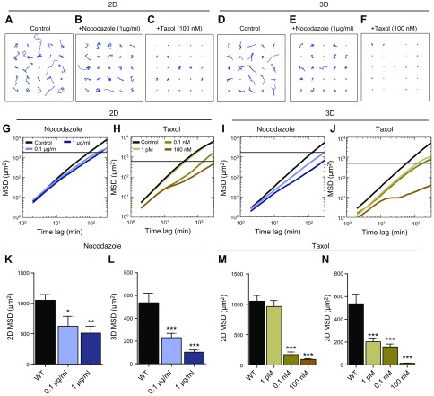
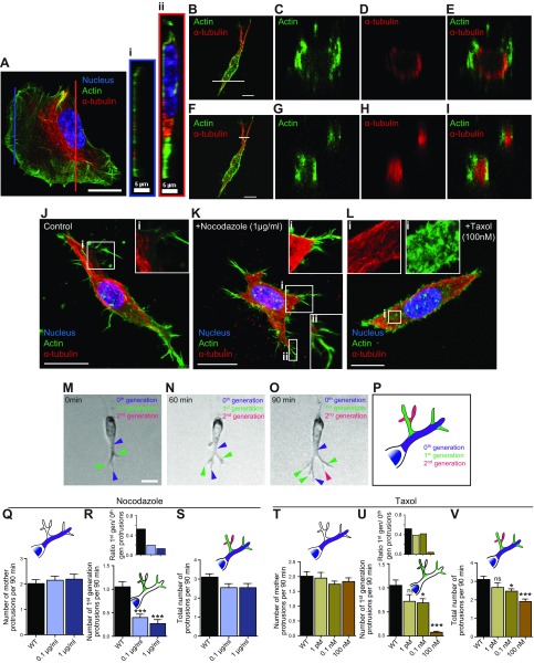
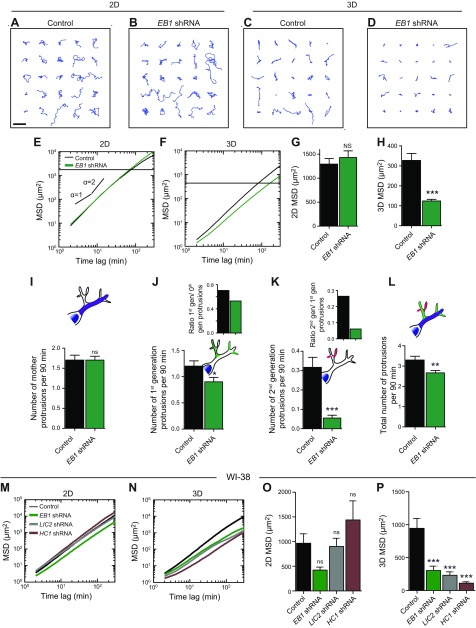
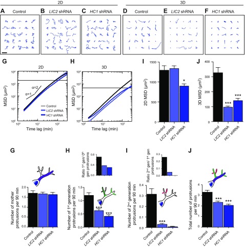
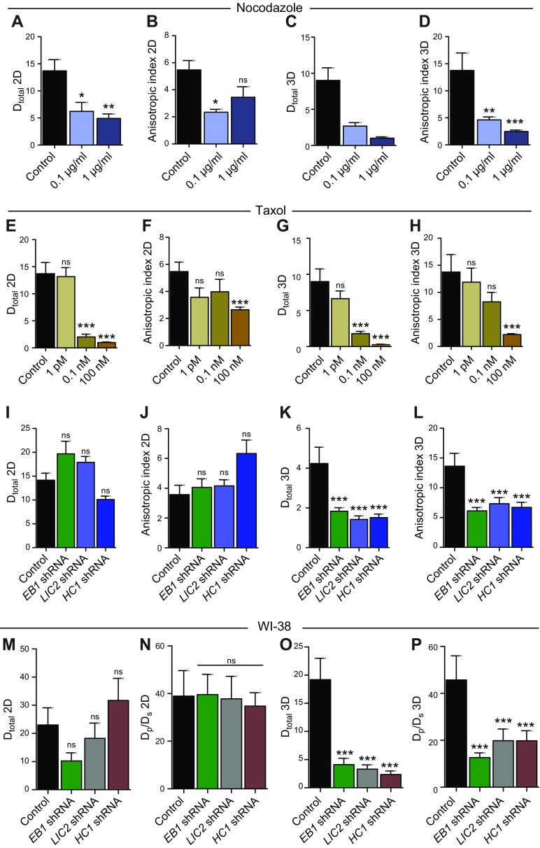
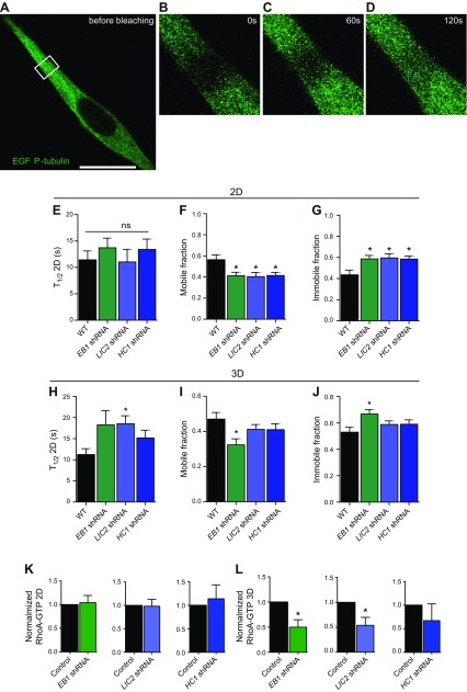
Microtubules have long been implicated to play an integral role in metastatic disease, for which a critical step is the local invasion of tumor cells into the 3-dimensional (3D) collagen-rich stromal matrix. Here we show that cell migration of human cancer cells uses the dynamic formation of highly branched protrusions that are composed of a microtubule core surrounded by cortical actin, a cytoskeletal organization that is absent in cells on 2-dimensional (2D) substrates. Microtubule plus-end tracking protein End-binding 1 and motor protein dynein subunits light intermediate chain 2 and heavy chain 1, which do not regulate 2D migration, critically modulate 3D migration by affecting RhoA and thus regulate protrusion branching through differential assembly dynamics of microtubules. An important consequence of this observation is that the commonly used cancer drug paclitaxel is 100-fold more effective at blocking migration in a 3D matrix than on a 2D matrix. This work reveals the central role that microtubule dynamics plays in powering cell migration in a more pathologically relevant setting and suggests further testing of therapeutics targeting microtubules to mitigate migration.-Jayatilaka, H., Giri, A., Karl, M., Aifuwa, I., Trenton, N. J., Phillip, J. M., Khatau, S., Wirtz, D. EB1 and cytoplasmic dynein mediate protrusion dynamics for efficient 3-dimensional cell migration.

Keywords: RhoA; microtubule; paclitaxel.


Conflict of interest statement

This work was supported by U.S. National Institutes of Health, National Cancer Institute Grants U54CA143868 and R01CA174388. The authors declare no conflicts of interest.

Figures

Figure 1.
Figure 1.
Microtubule dynamics mediates 3D cell migration. AF). Typical trajectories of 25 individual control HT-1080 cells and cells treated with the microtubule-depolymerizing drug nocadozole and the microtubule-stabilizing drug taxol, migrating on collagen I–coated 2D substrates and inside 3D collagen I matrices. G, H). Population-averaged MSDs of control cells and cells treated with nocodazole (G) or taxol (H) migrating on 2D substrates. I, J). Population-averaged MSDs of control cells and cells treated with nocodazole (I) or taxol (J) migrating inside 3D collagen I matrix. KN). MSDs of nocodazole- and taxol-treated cells migrating on 2D substrates (K, M) and in 3D matrix (L, M) evaluated at time scales of 1 h. For each condition, n = 3 biologic repeats; at least 60 cells were probed. *P < 0.05, **P < 0.01, ***P < 0.001.
Figure 2.
Figure 2.
Microtubule dynamics promotes protrusion branching in 3D matrix. AI). Actin filament and microtubule organization in control HT1080 cells growing on 2D substrates (A) and inside 3D matrix (BI). Cross-sections through the lamellipodium (right panel i, orange border) and the perinuclear cell body (right panel ii, red border). Cells were stained with DAPI (nuclear DNA) and antibodies against microtubule (red) and actin filament (green). JL). Immunofluorescent images of control cells (J) and cells treated with nocodazole (K) and taxol (L) embedded in 3D collagen I matrices. Insets: Daughter protrusions arising from the mother protrusions that prolong the nucleus are filled with microtubules in their lumen and F-actin at the periphery (J); nocodazole treatment increases the number of filopodial-like protrusions (K); taxol treatment gives rise to short, hairy actin protrusion throughout the cell (L). Cells were stained with DAPI (nuclear DNA) and antibodies against microtubule (red) and actin filament (green); images were obtained by immunofluorescence microscopy. MP). Active formation of pseudopodial protrusions by a cell embedded inside a 3D matrix (purple arrowheads, zeroth-generation or mother protrusions that stems directly from the cell body; green arrowheads, first-generation protrusions that start from the zeroth-generation protrusions; magenta arrowheads, second-generation protrusions that start from the first-generation protrusions). Schematic showing zeroth-, first-, and second-generation protrusions in a cell (P). QV). Total number of mother protrusions (zeroth-generation protrusions) generated per 90 min per cell in nocodazole- and taxol-treated cells (Q, T). Number of first-generation protrusions generated per 90 min per cell (R, U). Insets: Number of first-generation protrusions per mother protrusion (degree of branching) (R, U); total number of protrusions generated per 90 min per cell (S, V). For all panels, cells were monitored for 16.5 h. For each condition, n = 3; at least 40 cells were probed for protrusion topology analysis. WT, wild type. *P < 0.05, ***P < 0.001. Scale bars, 20 µm.
Figure 3.
Figure 3.
The distinct role of EB1 in 3D cell migration. AD). Typical trajectories of 25 individual control and EB1-depleted cells migrating on collagen I–coated 2D substrates and inside 3D collagen I matrices. Scale bar, 200 µm. E, F). shRNA-mediated depletion of EB1 has an inhibitory effect on cell migration in cells migrating in collagen I matrices (F) but has no significant effect on cell migration on substrates (E). G, H). Regulation of migration for cells on 2D substrates and embedded in 3D collagen I matrices by EB1. MSDs were evaluated at a time scale of 1 h. I). Total number of mother protrusions (zeroth-generation protrusions) generated per 90 min per cell. J, K). Number of first-generation protrusions (J) and second-generation protrusions (K) generated per 90 min per cell. Insets: Number of first-generation protrusions per mother protrusion (J); number of second-generation protrusions per first-generation protrusion (K). L). Total number of protrusions generated per 90 min per cell. For all panels, cells were monitored for 16.5 h. M, N). MSD plots for WI-38 cells depleted of EB1, LIC2, and HC1 moving on 2D substrates (M) and inside collagen I matrix (N). O, P). MSDs for WI-38 cells evaluated at a time scale of 1 h for cells on 2D substrates (O) and inside 3D matrix (P). For each condition, n = 3; at least 60 cells were probed for cell migration analysis, and at least 40 cells were probed for protrusion topology analysis. Ns, not significant. **P < 0.01, ***P < 0.001.
Figure 4.
Figure 4.
The distinct role of LIC2 and HC1 in 3D cell migration. AF). Typical trajectories of 25 individual control, LIC2-, and HC1-depleted cells migrating on collagen I–coated 2D substrates and inside 3D collagen I matrices. Scale bar, 200 µm. G, H). shRNA-mediated depletion of LIC2 has an inhibitory effect on cell migration in cells migrating in collagen I matrices (H) but has no significant effect on cell migration on substrates (G). I, J). Regulation of migration for cells on 2D substrates (I) and in 3D collagen I matrices (J) by LIC2. MSDs were evaluated at a time scale of 1 h (I, J). K). Total number of mother protrusions (zeroth-generation protrusions) generated per 90 min per cell. L, M). Number of first-generation protrusions (L) and second-generation protrusions (M) generated per 90 min per cell. Insets: Number of first-generation protrusions per mother protrusion (L); number of second-generation protrusions per first-generation protrusion (M). N). Total number of protrusions generated per 90 min per cell. For all panels, cells were monitored for 16.5 h. For each condition, n = 3; at least 60 cells were probed. *P < 0.05, ***P < 0.001.
Figure 5.
Figure 5.
Cell migration in 3D matrix is anisotropic. AD). Total diffusivity and anisotropic index for nocodazole-treated cells on 2D (A, B) and in 3D matrix (C, D). EH). Total diffusivity and anisotropic index for nocodazole-treated cells on 2D (E, F) and in 3D matrix (G, H). IL). Total diffusivity and anisotropic index for EB1-, LIC2-, and HC1-depleted cells on 2D (I, J) and in 3D matrix (K, L). MP). Total diffusivity and anisotropic index for EB1-, LIC2-, and HC1-depleted WI-38 cells on 2D (M, N) and in 3D matrix (O, P). Ns, not significant. *P < 0.05, **P < 0.01, ***P < 0.001.
Figure 6.
Figure 6.
LIC2 and EB1 promote fast microtubule dynamics in pseudopodial protrusions of cells in 3D matrix via RhoA. AD). FRAP images of cells embedded inside 3D collagen I matrices. Time of image acquisition is mentioned in the images. EJ). Half-life of fluorescence recovery, mobile fraction, and immobile fraction of α-tubulin GFP for control and for EB1-, LIC2-, and HC1-depleted cells on 2D substrates (EG) and inside a 3D matrix (HJ). K, L). RhoA-GTP. Ns, not significant. *P < 0.05.

References

    1. Ridley A. J., Schwartz M. A., Burridge K., Firtel R. A., Ginsberg M. H., Borisy G., Parsons J. T., Horwitz A. R. (2003) Cell migration: integrating signals from front to back. Science 302, 1704–1709 https://doi.org/10.1126/science.1092053 - DOI - PubMed
    1. Raftopoulou M., Hall A. (2004) Cell migration: rho GTPases lead the way. Dev. Biol. 265, 23–32 https://doi.org/10.1016/j.ydbio.2003.06.003 - DOI - PubMed
    1. Machesky L. M., Way M. (1998) Actin branches out. Nature 394, 125–126 https://doi.org/10.1038/28039 - DOI - PubMed
    1. Vladar E. K., Antic D., Axelrod J. D. (2009) Planar cell polarity signaling: the developing cell’s compass. Cold Spring Harb. Perspect. Biol. 1, a002964 https://doi.org/10.1101/cshperspect.a002964 - DOI - PMC - PubMed
    1. Giri A., Bajpai S., Trenton N., Jayatilaka H., Longmore G. D., Wirtz D. (2013) The Arp2/3 complex mediates multigeneration dendritic protrusions for efficient 3-dimensional cancer cell migration. FASEB J. 27, 4089–4099 - PMC - PubMed

Publication types

MeSH terms

Substances

LinkOut - more resources