. 2018 Feb;32(2):1025-1043.
doi: 10.1096/fj.201700182RRR. Epub 2018 Jan 3.

Ryanodine channel complex stabilizer compound S48168/ARM210 as a disease modifier in dystrophin-deficient mdx mice: proof-of-concept study and independent validation of efficacy

Affiliations

Ryanodine channel complex stabilizer compound S48168/ARM210 as a disease modifier in dystrophin-deficient mdx mice: proof-of-concept study and independent validation of efficacy

Roberta Francesca Capogrosso et al. FASEB J. 2018 Feb.

Abstract

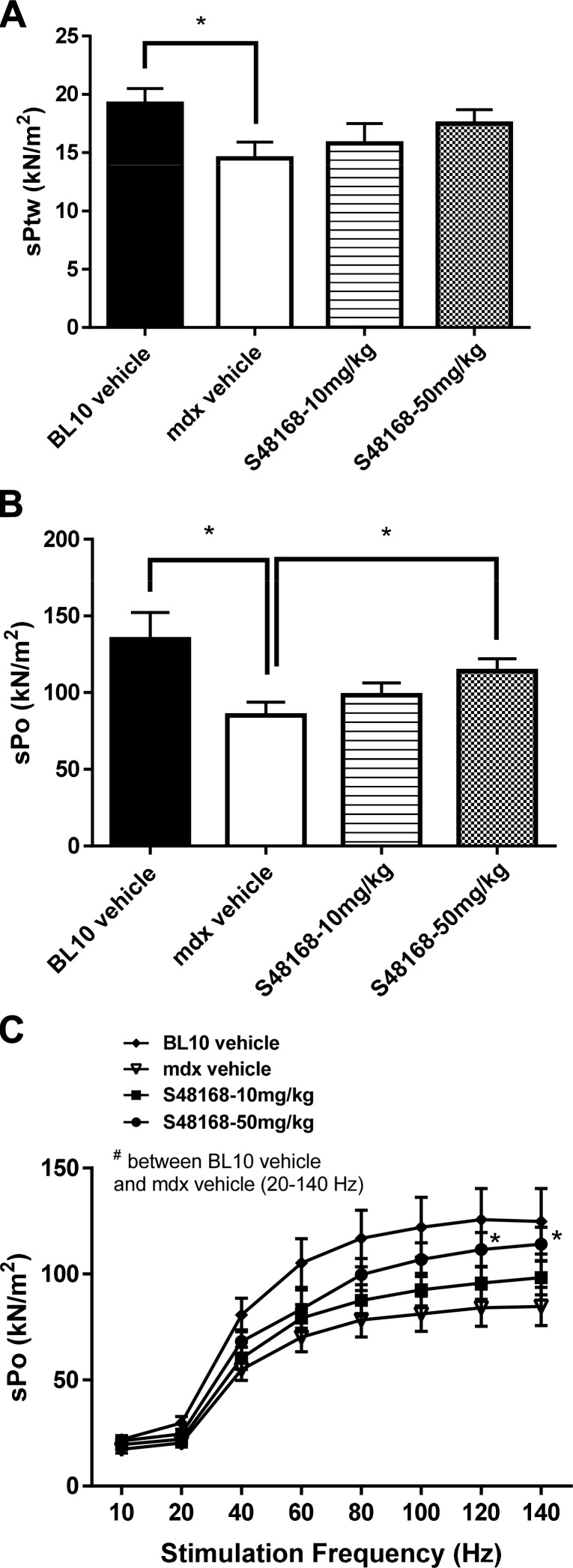
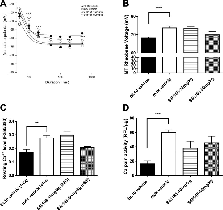
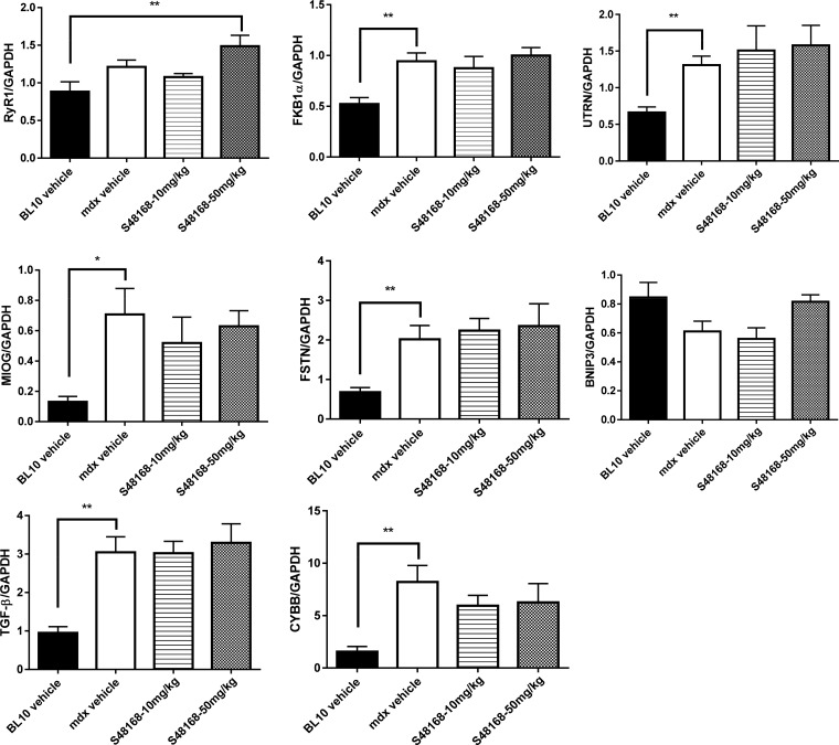
Muscle fibers lacking dystrophin undergo a long-term alteration of Ca2+ homeostasis, partially caused by a leaky Ca2+ release ryanodine (RyR) channel. S48168/ARM210, an RyR calcium release channel stabilizer (a Rycal compound), is expected to enhance the rebinding of calstabin to the RyR channel complex and possibly alleviate the pathologic Ca2+ leakage in dystrophin-deficient skeletal and cardiac muscle. This study systematically investigated the effect of S48168/ARM210 on the phenotype of mdx mice by means of a first proof-of-concept, short (4 wk), phase 1 treatment, followed by a 12-wk treatment (phase 2) performed in parallel by 2 independent laboratories. The mdx mice were treated with S48168/ARM210 at two different concentrations (50 or 10 mg/kg/d) in their drinking water for 4 and 12 wk, respectively. The mice were subjected to treadmill sessions twice per week (12 m/min for 30 min) to unmask the mild disease. This testing was followed by in vivo forelimb and hindlimb grip strength and fatigability measurement, ex vivo extensor digitorum longus (EDL) and diaphragm (DIA) force contraction measurement and histologic and biochemical analysis. The treatments resulted in functional (grip strength, ex vivo force production in DIA and EDL muscles) as well as histologic improvement after 4 and 12 wk, with no adverse effects. Furthermore, levels of cellular biomarkers of calcium homeostasis increased. Therefore, these data suggest that S48168/ARM210 may be a safe therapeutic option, at the dose levels tested, for the treatment of Duchenne muscular dystrophy (DMD).-Capogrosso, R. F., Mantuano, P., Uaesoontrachoon, K., Cozzoli, A., Giustino, A., Dow, T., Srinivassane, S., Filipovic, M., Bell, C., Vandermeulen, J., Massari, A. M., De Bellis, M., Conte, E., Pierno, S., Camerino, G. M., Liantonio, A., Nagaraju, K., De Luca, A. Ryanodine channel complex stabilizer compound S48168/ARM210 as a disease modifier in dystrophin-deficient mdx mice: proof-of-concept study and independent validation of efficacy.

Keywords: Duchenne muscular dystrophy; murine model; preclinical drug testing; rycals; skeletal muscle.


Conflict of interest statement

The authors thank Dr. Deborah McClellan for editing the manuscript. The compound used for this study was provided by Servier (Suresnes, France) and Armgo Pharma (Tarrytown, NY, USA), who are jointly developing S48168/ARM210 for indications including DMD. This study was supported in part by funds from Muscular Dystrophy Association Grant 228338 (Chicago, IL, USA); by U.S. National Institutes of Health Grant K26OD011171 (to K.N.); and by funds from the Dutch Duchenne Parent Project (NL-DPP) (to A.D.L.). K. N. and A. D. L. are senior coauthors. K. N. is a cofounder of Reveragen Biopharma, which is engaged in the development of therapeutic products for DMD. The remaining authors declare no conflicts of interest.

Figures

Figure 1.
Figure 1.
A, B) Body weight measurement (A) and mean water consumption normalized to body weight (B) over the 4 wk of treatment for WT mice, mdx mice treated with vehicle, and mdx mice treated with S48168/ARM210 (hereafter referred to as S48168) at 10 or 50 mg/kg per day. *P < 0.05; **P < 0.01, mdx vehicle-treated vs. WT mice for all time points except 2 wk after treatment. C) Calculated dose of S48168 received by mice at both concentrations (mg/kg) over the 4 wk of treatment is shown. DF) Baseline (t = 0) and posttreatment (t = 4) maximum forelimb grip strength (KGF; D), normalized forelimb strength to body weight (KGF/kg; E), and total distance run during an exhaustion test (F) for all experimental groups. All values are expressed as means ± sem (n = 6–10 animals/group). *P < 0.05; **P < 0.01; ***P < 0.001, WT vs. mdx vehicle-treated mice, by unpaired Student’s t test, and mdx vehicle-treated vs. S48168-treated groups, by 1-way ANOVA with Dunnett’s multiple comparison test, as indicated. Although not shown, there was a statistically significant difference between the mdx vehicle-treated and WT mice in (F).
Figure 2.
Figure 2.
Specific twitch force (sPtw; A), specific tetanic force (sPo; B), and force–frequency curves (C) for DIA muscles in WT mice, mdx mice treated with vehicle, and mdx mice treated with S48168 at 10 or 50 mg/kg per day, after 4 wk of treatment. All values are expressed as means ± sem (n = 6–10/group). *P < 0.05, WT vs. mdx vehicle-treated mice, by unpaired Student’s t test, and mdx vehicle-treated vs. S48168-treated mice at 50 mg/kg per day, by 1-way ANOVA with Dunnett’s multiple comparison test.
Figure 3.
Figure 3.
A, B) Effect of 4 wk of treatment with S48168 on the mechanical threshold (MT) of mdx EDL muscle fibers; the curves for membrane voltages for myofiber contraction in relation to the duration of the depolarizing pulse (strength–duration curves) are shown in A. The MT rheobase voltage values obtained from the fit of the curves are shown in BD, which show the resting intracellular Ca2+ concentration of FDB muscle fibers and calpain activity in DIA, respectively. Each graph shows the values from WT mice, mdx mice treated with vehicle, and mdx mice treated with S48168 at 10 or 50 mg/kg per day. AC) Data are expressed as means ± sem from 40 to 11 values (n = 4–5 mice. D) All values are expressed as means ± sem from 5 animals per group. BD) **P < 0.01; ***P < 0.001, WT vs. mdx vehicle-treated group, by unpaired Student’s t test.
Figure 4.
Figure 4.
Representative images of H&E-stained DIA and GC muscles, exhibiting mild-to-severe phenotypic variances of mdx mice treated with vehicle or with S48168 at 10 or 50 mg/kg per day after 4 wk of treatment. Scale bars, 200 µm.
Figure 5.
Figure 5.
Area of total damage and regeneration in GC muscle (A) and DIA (B) from mdx mice treated with vehicle or S48168 at 10 or 50 mg/kg per day after 4 wk of treatment. All values are expressed as means ± sem (n = 6 animals per group). *P < 0.05; ***P < 0.001, vehicle- vs. S48168-treated mice, by 1-way ANOVA with Dunnett’s multiple comparison test.
Figure 6.
Figure 6.
Gene expression profiling of RyR1, FKB1α, UTRN, MIOG, FSTN, BNIP3, TGF-β, and CYBB normalized to GAPDH for WT mice, mdx mice treated with vehicle, or mdx mice treated with S48168 at 10 or 50 mg/kg per day after 4 wk of treatment. All values are expressed as means ± sem (n = 6 animals per group). *P < 0.05; **P < 0.01 WT vs. mdx vehicle-treated mice, by unpaired Student’s t test, and WT mice vs. mdx treated mice, by 1-way ANOVA with Dunnett’s multiple comparison test between as indicated.
Figure 7.
Figure 7.
Body weight measurements from UNIBA (A) and AGA (D) and mean water consumption normalized to body weight from UNIBA (B) and AGA (E) over the 12 wk of treatment for WT mice, mdx mice treated with vehicle, and mdx mice treated with S48168 at 10 or 50 mg/kg per day. The calculated doses of S48168 received by the mice at both concentrations (mg/kg per day) over the 12 wk of treatment are shown in UNIBA (C) and AGA (F) All values are expressed as means ± sem (n = 8 to 15 animals per group).
Figure 8.
Figure 8.
Representative images of H&E-stained DIA and GC muscles exhibiting mild-to-severe phenotypic variations in mdx mice treated with vehicle or with S48168 at10 or 50 mg/kg per day for 12 wk. Scale bar, 200 µm.

References

    1. Roses A. D., Herbstreith M. H., Appel S. H. (1975) Membrane protein kinase alteration in Duchenne muscular dystrophy. Nature 254, 350–351 - PubMed
    1. Hoffman E. P., Schwartz L. (1991) Dystrophin and disease. Mol. Aspects Med. 12, 175–194 - PubMed
    1. Hoffman E. P., Brown R. H. Jr., Kunkel L. M. (1987) Dystrophin: the protein product of the Duchenne muscular dystrophy locus. Cell 51, 919–928 - PubMed
    1. Ervasti J. M., Ohlendieck K., Kahl S. D., Gaver M. G., Campbell K. P. (1990) Deficiency of a glycoprotein component of the dystrophin complex in dystrophic muscle. Nature 345, 315–319 - PubMed
    1. Ohlendieck K., Campbell K. P. (1991) Dystrophin-associated proteins are greatly reduced in skeletal muscle from mdx mice. J. Cell Biol. 115, 1685–1694 - PMC - PubMed

Publication types

MeSH terms