. 2017 Nov 3;358(6363):eaao0464.
doi: 10.1126/science.aao0464.

Structure of the mitochondrial inner membrane AAA+ protease YME1 gives insight into substrate processing

Affiliations

Structure of the mitochondrial inner membrane AAA+ protease YME1 gives insight into substrate processing

Cristina Puchades et al. Science. .

Abstract

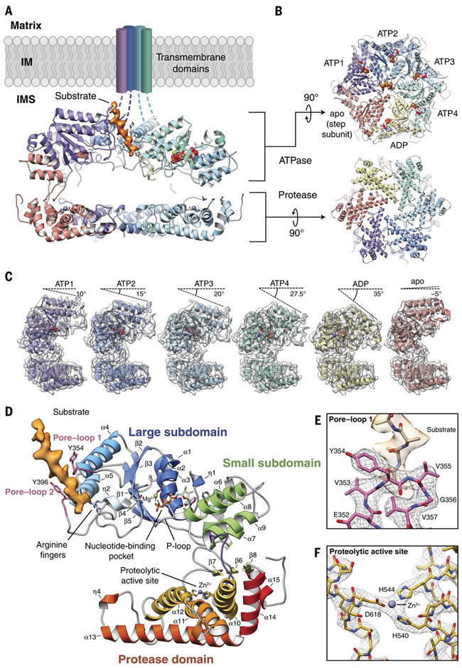
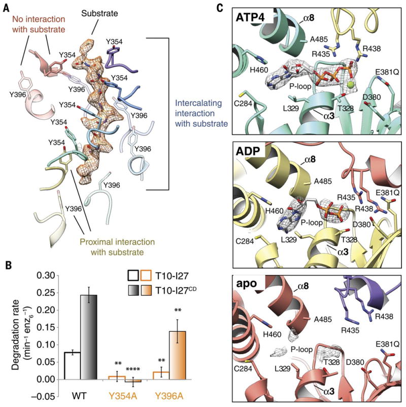
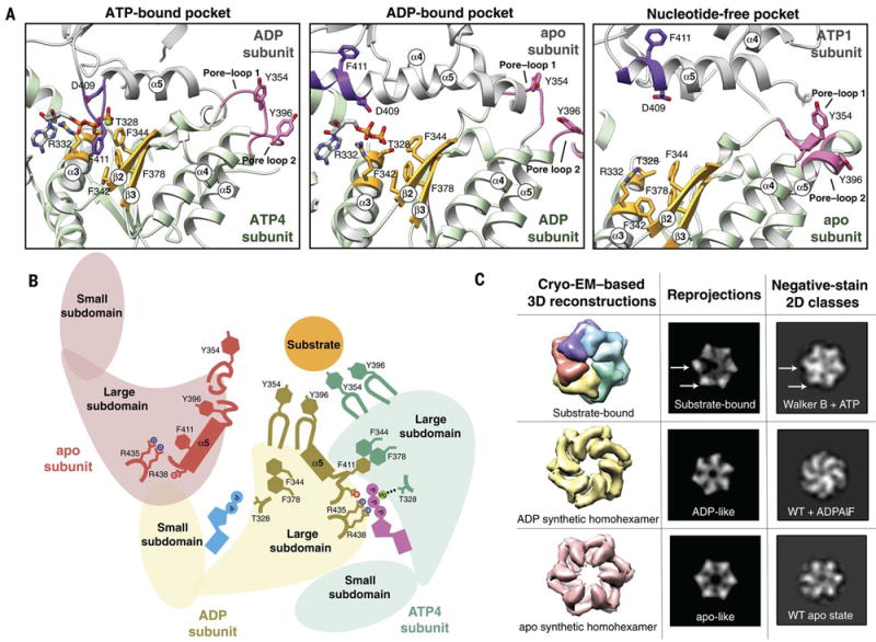
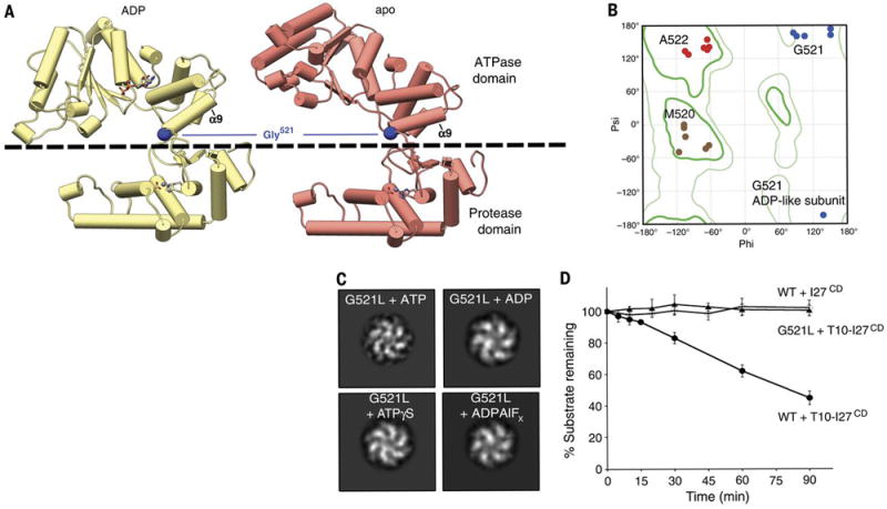
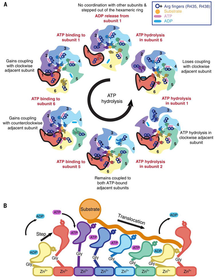
We present an atomic model of a substrate-bound inner mitochondrial membrane AAA+ quality control protease in yeast, YME1. Our ~3.4-angstrom cryo-electron microscopy structure reveals how the adenosine triphosphatases (ATPases) form a closed spiral staircase encircling an unfolded substrate, directing it toward the flat, symmetric protease ring. Three coexisting nucleotide states allosterically induce distinct positioning of tyrosines in the central channel, resulting in substrate engagement and translocation to the negatively charged proteolytic chamber. This tight coordination by a network of conserved residues defines a sequential, around-the-ring adenosine triphosphate hydrolysis cycle that results in stepwise substrate translocation. A hingelike linker accommodates the large-scale nucleotide-driven motions of the ATPase spiral relative to the planar proteolytic base. The translocation mechanism is likely conserved for other AAA+ ATPases.


Figures

Fig. 1
Fig. 1. Architecture of the substrate-bound YME1 AAA+ protease
A) Cutaway view of the substrate-bound YME1 atomic model, with cryo-EM density for substrate colored orange. Each subunit of the homohexamer is assigned a unique color and the nucleic acids are depicted using a sphere representation in (A-C). B) Views of the ATPase and protease rings, shown orthogonally to the orientation shown in (A). C) Individual protomers shown side-by-side, aligned with the protease domain in the same orientation. The cryo-EM density is shown as a transparent gray isosurface, showing the quality of the reconstruction. The sequential movement of the ATPase domain relative to the protease domain is emphasized by a dashed line above each protomer, depicting the tilt of the pore loop helices relative to the horizontal protease ring. The pore loop tyrosines are visible within each of the pore loops. D) Topological organization of the YME1 protomer showing the large and small subdomains of the ATPase domain, and protease domain underneath. Notable and conserved components of the YME1 subunit are highlighted. The cryo-EM density of the substrate is shown in orange. E) Detailed view of the pore loop 1 interactions with substrate. The cryo-EM density of the substrate is shown as a transparent orange isosurface, along with a poly-alanine model of the substrate. The cryo-EM density of the pore loop is shown as a mesh, and the atomic model shows the interaction of Tyr354 and Val355 with substrate. F) Detailed view of the zinc-coordinated proteolytic active site.
Fig. 2
Fig. 2. Nucleotide state correlates with pore loop conformation and substrate interaction
A) Three categories of interactions are observed between the pore loop tyrosines and substrate. A poly-alanine model of the substrate is shown within the cryo-EM density of the substrate, shown as orange mesh. Tyr354 and Tyr396 from pore loops 1 and 2 are rendered as solid and semi-transparent, respectively. The Tyr residues from the ATP(1-4) subunits (purple, blue, cyan, and turquoise) show close, stable, intercalating interactions with substrate, while Tyr residues from the ADP subunit (yellow) are positioned further from the substrate, and Tyr residues from the apo subunit (red) show no interaction with substrate. B) Effect of mutations in the conserved pore loops on substrate degradation. Initial degradation rates are shown for folded (T10-I27) and unfolded (T10-I27CD) substrates by wild-type hexYME1 and variants bearing either Y354A or Y396A substitutions. Mutation of the pore loop 1 Tyrosine abolishes degradation for both substrates and mutation of the pore loop 2 Tyrosine significantly diminishes the degradation rate for both substrates, consistent with an important role for both of these residues in substrate handling. Values are means of independent replicates (n≥3) ± s.d. **P≤0.01, ****P<0.0001 as calculated using the Student’s two-tailed t-test and shown in comparison to the degradation of the identical substrate by wild-type hexYME1. C) Organization of the nucleotide binding pockets in the ATP(1-4), ADP, and apo states. Cryo-EM density corresponding to the nucleotide (or lack thereof) is shown as a gray mesh at a σ level of 4.0 for all states. Absence of gamma phosphate and magnesium ion is evident in the ADP-like state, whereas only a low level of nucleotide density is still present in the apo-conformation (bottom panel). In the ATP(1-4) and ADP conformations, the adenine base is located within 4 Å of L329 and C284 of the large domain and H460 of the small domain, whereas the ribose directly interacts with the backbone of A485 from helix α8 of the small domain. The arginine fingers (R435 and R438) are closely coordinated with the phosphates in the ATP(1-4) subunits, but further away in the ADP and apo subunits.
Fig. 3
Fig. 3. Nucleotide state causes major conformational rearrangements of the ATPase domain
A) The three nucleotide state conformations were aligned to the central phenylalanine-containing β-sheet (colored gold) to show the major rearrangements that allosterically affect pore loop conformation. In the ATP-bound pocket, the ISS loop of the adjacent subunit (colored purple) extends towards the Phe-containing β-sheet. In this conformation, the pore loops (colored pink) are extended toward the central pore and interact strongly with substrate. In the ADP-bound pocket, loss of the gamma phosphate results in a retraction of the ISS loop from the Phe-containing β-sheet, and the ISS loop becomes part of the α5 helix of the adjacent subunit. Within the ADP-bound subunit, the α3 helix moves closer to the Phe-containing β-sheet, and the pore-loop tyrosines more weakly interact with substrate. In the nucleotide-free state, the pore loop tyrosines are completely dissociated from the substrate, and are incorporated into helices 4 and 5. B) Cartoon representation of the nucleotide-dependent conformational changes affecting the entire ATPase domain, depicting the motions described in (A). When ATP is bound within the nucleotide pocket, the pore loop tyrosines interact strongly with substrate. ATP hydrolysis results in a weakening of the substrate-tyrosine interactions, and loss of the nucleotide results in a complete release of substrate. C) Nucleotide-state is shown to cause major domain rotations, as 3D structures of the substrate-bound and computationally symmetrized ADP and apo homohexamers generate distinctly different 2D projections that show structural characteristics that are similar to those in reference-free 2D classes obtained by negative stain EM in the presence of ATP, ADPALFX or in the absence of nucleotide. Wild type hexYME1 is abbreviated as WT.
Fig. 4
Fig. 4. A hinge-like glycine linker accommodates nucleotide-state induced domain rotations
A) The ADP and apo subunits are shown (yellow and red, respectively), aligned relative to their protease domains. The dashed line delineates the ATPase domain (above) from the protease domain (below). The ATPase domain undergoes a large movement as it progresses from the ADP to the nucleotide-free state, which stems from a pivoting of the α9 helix around the inter-domain linker. Gly521 within this linker is shown to play a key role in enabling this large-scale motion. B) Ramachandran plot displaying phi and psi angles of inter-domain residues M520 (brown), G521 (blue) and A522 (red) in each subunit shows major torsion changes of G521 in the ADP-like state. The location of the Gly521 phi and psi angles underscore the necessity of a torsionally flexible Gly in this position. C) 2D image analyses of a negatively stained hexYME1 construct containing a G521L mutation show that addition of ATP, ADP, ADPALFX, and ATPγS do not significantly influence the quaternary organization of the complex. Since all the resulting class averages appear to have a similar organization as the ADP-like conformation shown in Fig. 3C, we suspect that the subunits of this mutant construct are trapped in the ADP conformation. D) Plot showing the effect of the G521L mutation on the degradation of an unfolded substrate. Rapid loss of T10-I27CD is seen over time in the presence of wild-type hexYME1 whereas no loss of T10-I27CD is observed in the presence of the G521L point mutant or for unfolded I27CD lacking the T10 degron incubated with wild-type hexYME1. Values are means of independent replicates (n≥3) ± s.d.
Fig. 5
Fig. 5. Model for ATP-driven substrate translocation into the proteolytic chamber of YME1
A) Cartoon representation of the ATP hydrolysis cycle that proceeds sequentially in a counterclockwise manner around the hexameric ATPase ring. We show how the coordinated nucleotide-dependent changes in the hexamer affect the subunit shown in red throughout the cycle. ATP and ADP are represented as blue and pink ovals at the interprotomer interface, and an orange circle depicts the substrate trapped in the central pore and coordinated by the pore-loops. B) Scheme of the progressive conformational rearrangements of a given subunit as it progresses through a single ATP-hydrolysis cycle (from left to right): ADP release, ATP-binding, ATP-bound states, ATP-hydrolysis, ADP-state, and ADP release. Each nucleotide state is highlighted by a different color. The yellow and red subunits are shown twice to highlight the complete cycle.

References

    1. Quiros PM, Langer T, LopezOtin C. New roles for mitochondrial proteases in health, ageing and disease. Nat Rev Mol Cell Biol. 2015;16:345–359. - PubMed
    1. Tatsuta T, Langer T. Quality control of mitochondria: protection against neurodegeneration and ageing. EMBO J. 2008;27:306–314. - PMC - PubMed
    1. Nolden M, et al. The m-AAA protease defective in hereditary spastic paraplegia controls ribosome assembly in mitochondria. Cell. 2005;123:277–289. - PubMed
    1. Baker MJ, Tatsuta T, Langer T. Quality control of mitochondrial proteostasis. Cold Spring Harb Perspect Biol. 2011;3 - PMC - PubMed
    1. Leonhard K, et al. AAA proteases with catalytic sites on opposite membrane surfaces comprise a proteolytic system for the ATP-dependent degradation of inner membrane proteins in mitochondria. EMBO J. 1996;15:4218–4229. - PMC - PubMed

Publication types

MeSH terms

Substances

LinkOut - more resources