. 2017 Nov 3;358(6363):668-672.
doi: 10.1126/science.aan0092.

Spindle asymmetry drives non-Mendelian chromosome segregation

Affiliations

Spindle asymmetry drives non-Mendelian chromosome segregation

Takashi Akera et al. Science. .

Abstract

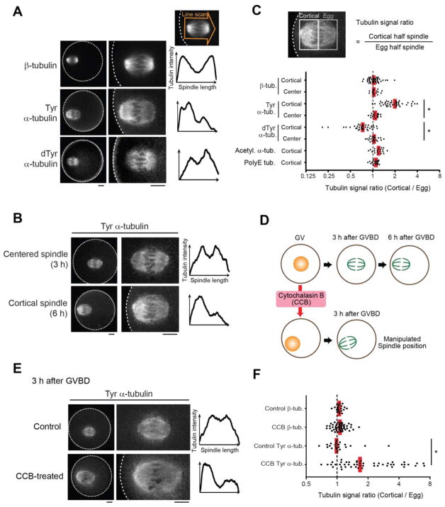
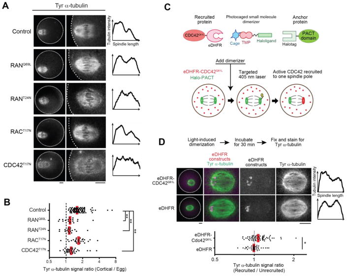
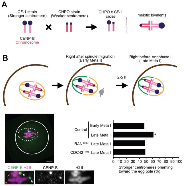
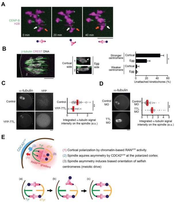
Genetic elements compete for transmission through meiosis, when haploid gametes are created from a diploid parent. Selfish elements can enhance their transmission through a process known as meiotic drive. In female meiosis, selfish elements drive by preferentially attaching to the egg side of the spindle. This implies some asymmetry between the two sides of the spindle, but the molecular mechanisms underlying spindle asymmetry are unknown. Here we found that CDC42 signaling from the cell cortex regulated microtubule tyrosination to induce spindle asymmetry and that non-Mendelian segregation depended on this asymmetry. Cortical CDC42 depends on polarization directed by chromosomes, which are positioned near the cortex to allow the asymmetric cell division. Thus, selfish meiotic drivers exploit the asymmetry inherent in female meiosis to bias their transmission.


Figures

Fig. 1
Fig. 1. Cortical proximity induces asymmetry within the mouse oocyte spindle
(A–F) CF-1 oocytes were fixed at metaphase I and stained for the indicated post-translational modifications on tubulin. Cortical spindles (A–C) were examined at 6 h after GVBD, and centered spindles (B, C) at 3 h after GVBD. Cortical spindles in oocytes treated with cytochalasin B were examined at 3 h after GVBD (D–F). Images (A, B, E) are sum intensity z-projections showing the whole oocyte (left) or a magnified view of the spindle (right); dashed line, cortex; scale bars, 10 μm. Graphs are line scans of tubulin intensity across the spindle. Spindle asymmetry was quantified (C, F) as the ratio of the cortical half to the egg half (n > 18 spindles for each condition). Each dot represents a single spindle; red line, median; *p < 0.0001.
Fig. 2
Fig. 2. Cortical polarization and localized CDC42 signaling induce spindle asymmetry
(A, B) CF-1 oocytes expressing the indicated GTPase mutant were fixed 6 h after GVBD and stained for Tyr α-tubulin. Images are sum intensity z-projections showing the whole oocyte (left) or a magnified view of the spindle (right), and graphs are line scans of tubulin intensity across the spindle. Spindle asymmetry was quantified (B) as the ratio of the cortical half to the egg half (n > 17 spindles for each condition). (C) Schematics of the light-induced dimerization experiment. The dimerizer is composed of a Halo ligand linked to the eDHFR ligand Trimethoprim (TMP), which is photocaged. The PACT domain, fused to EGFP and Halotag, localizes to spindle poles, and CDC42Q61LΔCAAX is fused to mCherry and eDHFR. The dimerizer covalently binds Halo-PACT at spindle poles, and eDHFR-CDC42Q61LΔCAAX is recruited to one pole by local uncaging with light. (D) Halo-EGFP-PACT was co-expressed with either mCherry-eDHFR-CDC42Q61LΔCAAX (top) or mCherry-eDHFR (bottom) in CF-1 oocytes. Recruitment of eDHFR fusion proteins was induced by uncaging at one spindle pole. 30 min after uncaging, oocytes were fixed and stained for Tyr α-tubulin. Images are maximum intensity z-projection showing whole oocytes (left) or magnified views of the spindle, and graphs are line scans of tubulin intensity across the spindle. Spindle asymmetry was quantified as the ratio of the recruited side to the unrecruited side (n > 39 spindles for each condition). Each dot represents a single spindle; red line, median; *p < 0.01; **p < 0.0001. Scale bars, 10 μm.
Fig. 3
Fig. 3. Spindle asymmetry is essential for biased orientation of selfish centromeres
(A) Schematic of biased orientation assay. A strain with stronger centromeres (CF-1) is crossed to a strain with weaker centromeres (CHPO). Bivalents in the hybrid offspring contain both stronger and weaker centromeres, which can be distinguished by CENP-B levels. (B) CHPO x CF-1 hybrid oocytes expressing CENP-B-EGFP and H2B-mCherry were imaged live, either shortly after spindle migration to the cortex (within 30 min, early meta I), or shortly before anaphase onset (within 30 min, late meta I). Image is a maximum intensity z-projection showing late meta I; white line: oocyte cortex, dashed line: spindle outline; scale bar, 10 μm. Insets are optical slices showing two bivalents; arrows indicate stronger (white) and weaker (orange) centromeres. The fraction of bivalents with the stronger centromere oriented towards the egg was quantified; n=152 bivalents for early meta I, 204 for late meta I, 108 for RanQ69L and 143 for CDC42T17N. * indicates significant deviation from 50% (p < 0.005).
Fig. 4
Fig. 4. MT tyrosination promotes unstable interactions between selfish centromeres and the cortical side of the spindle
(A) CHPO x CF-1 oocytes expressing CENP-B-mCherry and H2B-EGFP were imaged live after spindle migration to the cortex (n = 23 cells). Time lapse images show an example of bivalent flipping; arrows indicate stronger (white) and weaker (orange) centromeres. (B) CHPO x CF-1 oocytes were analyzed for cold-stable MTs at 8 h after GVBD. Enlarged insets are optical slices showing individual bivalents with the stronger centromere (arrow) either facing the egg side and attached to cold-stable MTs (1) or facing the cortical side and not attached (2). Weaker centromeres are attached in both cases. Graph shows the average percentage of centromeres without cold-stable attachments. Error bars represent s.d. for 3 independent experiments (> 50 bivalents analyzed in each experiment). *p < 0.01. (C, D) CF-1 oocytes expressing YFP-TTL or microinjected with morpholino against TTL were analyzed for cold-stable MTs at 6 h after GVBD. Graphs show integrated α-tubulin intensity in the spindle (n > 41 spindles for each condition). Each dot represents a single spindle; red line, median; *p < 0.001. Images (A–D) are maximum intensity z-projections; scale bars, 10 μm. (E) Model for spindle asymmetry and meiotic drive. Top: cortical signals regulate MTs to induce tyrosination asymmetry within the spindle, and stronger centromeres (larger blue circles) orient preferentially to the egg side. Bottom: bivalent orientation is initially random (a), but attachment of a stronger centromere to the cortical side is unstable and tends to detach (b), followed by detachment of the weaker centromere, likely due to loss of tension across the bivalent, and re-orientation. This biased flipping of stronger centromeres to the egg side leads to biased orientation (c).

Comment in

References

    1. Werren JH. Selfish genetic elements, genetic conflict, and evolutionary innovation. Proc Natl Acad Sci U S A. 2011;108:10863–70. - PMC - PubMed
    1. Rice WR. Nothing in Genetics Makes Sense Except in Light of Genomic Conflict. Annu Rev Ecol Evol Syst. 2013;44:217–237.
    1. Lindholm AK, et al. The Ecology and Evolutionary Dynamics of Meiotic Drive. Trends Ecol Evol. 2016;31:315–326. - PubMed
    1. Helleu Q, Gérard PR, Montchamp-Moreau C. Sex chromosome drive. Cold Spring Harb Perspect Biol. 2014;7:a017616. - PMC - PubMed
    1. Pardo-Manuel de Villena F, Sapienza C. Nonrandom segregation during meiosis: the unfairness of females. Mamm Genome. 2001;12:331–9. - PubMed

Publication types