Review
. 2017 Dec 4;216(12):3931-3948.
doi: 10.1083/jcb.201701095. Epub 2017 Nov 2.

How to rewire the host cell: A home improvement guide for intracellular bacteria

Affiliations
Review

How to rewire the host cell: A home improvement guide for intracellular bacteria

Elias Cornejo et al. J Cell Biol. .

Abstract

Intracellular bacterial pathogens have developed versatile strategies to generate niches inside the eukaryotic cells that allow them to survive and proliferate. Making a home inside the host offers many advantages; however, intracellular bacteria must also overcome many challenges, such as disarming innate immune signaling and accessing host nutrient supplies. Gaining entry into the cell and avoiding degradation is only the beginning of a successful intracellular lifestyle. To establish these replicative niches, intracellular pathogens secrete various virulence proteins, called effectors, to manipulate host cell signaling pathways and subvert host defense mechanisms. Many effectors mimic host enzymes, whereas others perform entirely novel enzymatic functions. A large volume of work has been done to understand how intracellular bacteria manipulate membrane trafficking pathways. In this review, we focus on how intracellular bacterial pathogens target innate immune signaling, the unfolded protein response, autophagy, and cellular metabolism and exploit these pathways to their advantage. We also discuss how bacterial pathogens can alter host gene expression by directly modifying histones or hijacking the ubiquitination machinery to take control of several host signaling pathways.


Figures

Figure 1.
Figure 1.
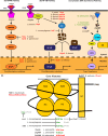
Modulation of innate immunity signaling pathways by bacterial pathogens. (A) Stimulation of PRRs by PAMPs activates a MAPK signaling cascade. (B) PRR activation also releases NF-κB from its inhibitor, IκB, which allows NF-κB to translocate to the nucleus and induce the expression of proinflammatory cytokines. (C) STING, a major regulator of the CSP that is anchored to the ER, is activated by cyclic dinucleotides cGAMP produced by DNA sensing from cGAS as well as secreted c-di-AMP produced by bacteria. Activation of the STING/TBK1/IRF3 pathway leads to a type I IFN response. (D) Posttranslational modifications to histones by bacterial pathogens. Bacterial effectors can inhibit host-mediated histone modifications by indirectly causing the reversal of these modifications. Bacterial effectors can also directly modify histones. H, histones; red, inhibitory histone modifications; green, activating histone modifications; black, pathogen effector protein; italics, pathogen. Histone modifications: phosphorylation (p), acetylation (ac), methylation (me), dimethylation (me2), and trimethylation (me3). Bacteria or secreted bacterial effectors can either inhibit (red) or activate (green) these innate immune signaling pathways.
Figure 2.
Figure 2.
Modulation of the UPR by bacterial pathogens. The UPR is mediated by three major sensors in the ER: IRE1, PERK, and ATF6. In the presence of unfolded proteins, the ER resident chaperone, BiP, dissociates from these UPR sensors, which contributes to their activation and downstream cellular responses, which include expression of protein chaperones and ERAD. Bacterial pathogens both activate (green) and inhibit (red) all three branches of the UPR.
Figure 3.
Figure 3.
Modulation of the autophagy pathway by bacterial pathogens. After invasion of the host cell, vacuoles containing intracellular bacteria are targeted for the autophagy machinery by ubiquitination. Adapter proteins specifically direct ubiquitinated bacteria containing vacuoles to LC3–PE conjugates on mature isolation membranes. The membrane expands and forms a double-membrane compartment called an autophagosome, which eventually fuses with lysosomes and leads to the degradation of its bacterial cargo. Several bacteria have evolved different effector proteins that inhibit (red) this selective autophagic immune mechanism. However, some bacterial pathogens secrete effectors that exploit this pathway (green) and make use of the generated nutrients and membranes to promote their intracellular growth.
Figure 4.
Figure 4.
Modulation of mTOR signaling during infection. During steady-state conditions, mTOR is a negative regulator of autophagy and inflammation, as well as a positive regulator of de novo lipogenesis (black lines). (A) During infection, the host will inhibit mTOR signaling to activate autophagy and inflammation and inhibit de novo lipogenesis. It does so by targeting mTOR and positive regulators of mTOR (i.e., AKT and PI3K) for proteasomal degradation. mTOR is also inhibited by amino acid starvation that occurs during infection by certain pathogens (e.g., S. typhimurium and S. flexneri). Some bacterial pathogens (e.g., S. typhimurium or L. pneumophila) activate positive regulators of mTOR signaling to counteract these host-driven effects and thereby improve their intracellular housing capacity. In the case of S. typhimurium, AKT is activated by FAK that is recruited to the surface of Salmonella-containing vacuoles.
Figure 5.
Figure 5.
Ubiquitination of host proteins by Legionella SidE effector family. The SidE family of effectors from L. pneumophila modify host ubiquitin and ubiquitinate host proteins using a novel catalytic mechanism. The DUB domain of SidE effectors does not interfere with SidE-mediated ubiquitination, but instead, removes ubiquitin imparted by the canonical ubiquitination machinery of the host. It is unclear whether the DUB activity acts to generate a pool of ubiquitin and/or a pool of host target protein substrates for SidE effectors. The mono-ADP-ribosyltransferase (mART) domain uses NAD+ to attach a phosphoribose moiety to arginine 42 of host ubiquitin, which generates ADP-ribosylated ubiquitin (ADPR-Ub) and nicotinamide (NAA). ADPR-Ub is further cleaved into phosphoribosylated ubiquitin (PR-Ub) and AMP by the nucleotidase/phosphohydrolase/phosphodiesterase (NP/PDE) domain. PR-Ub is covalently attached to host proteins via a noncanonical serine-linked phosphodiester bond. This novel ubiquitination mechanism does not require E1, E2, or E3 enzymes or ATP from the host. The generated pool of PR-Ub also disrupts canonical host ubiquitination machinery.

References

    1. Abe T., and Barber G.N.. 2014. Cytosolic-DNA-mediated, STING-dependent proinflammatory gene induction necessitates canonical NF-κB activation through TBK1. J. Virol. 88:5328–5341. 10.1128/JVI.00037-14 - DOI - PMC - PubMed
    1. Abshire C.F., Dragoi A.-M., Roy C.R., and Ivanov S.S.. 2016. MTOR-driven metabolic reprogramming regulates Legionella pneumophila intracellular niche homeostasis. PLoS Pathog. 12:e1006088 10.1371/journal.ppat.1006088 - DOI - PMC - PubMed
    1. Adachi Y., Yamamoto K., Okada T., Yoshida H., Harada A., and Mori K.. 2008. ATF6 is a transcription factor specializing in the regulation of quality control proteins in the endoplasmic reticulum. Cell Struct. Funct. 33:75–89. 10.1247/csf.07044 - DOI - PubMed
    1. Akira S., Uematsu S., and Takeuchi O.. 2006. Pathogen recognition and innate immunity. Cell. 124:783–801. 10.1016/j.cell.2006.02.015 - DOI - PubMed
    1. Alix E., Mukherjee S., and Roy C.R.. 2011. Subversion of membrane transport pathways by vacuolar pathogens. J. Cell Biol. 195:943–952. 10.1083/jcb.201105019 - DOI - PMC - PubMed

Publication types

MeSH terms