. 2017 Nov 2;7(1):14972.
doi: 10.1038/s41598-017-14966-w.

TDP-43 accelerates age-dependent degeneration of interneurons

Affiliations

TDP-43 accelerates age-dependent degeneration of interneurons

Hitomi Tsuiji et al. Sci Rep. .

Abstract

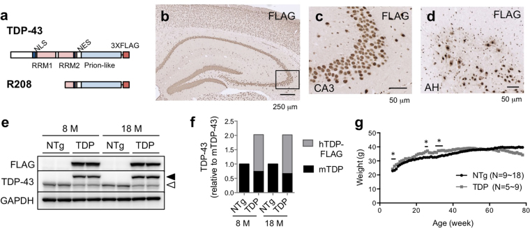
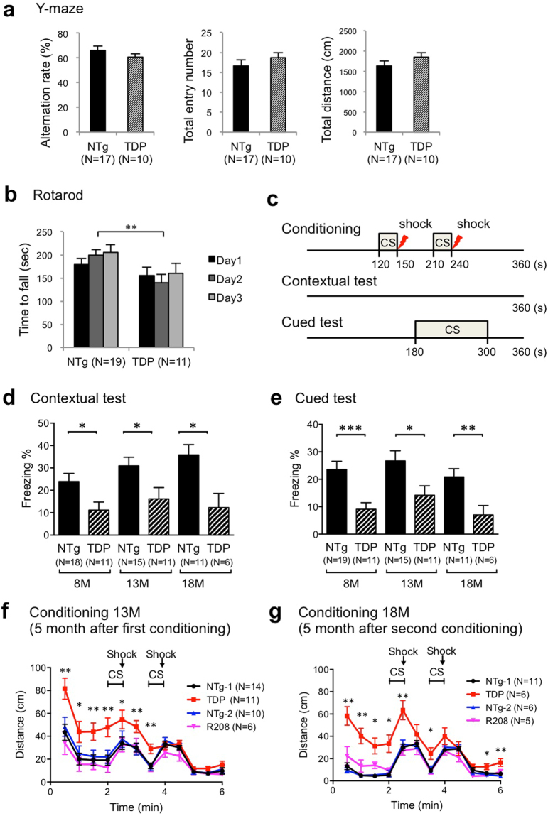
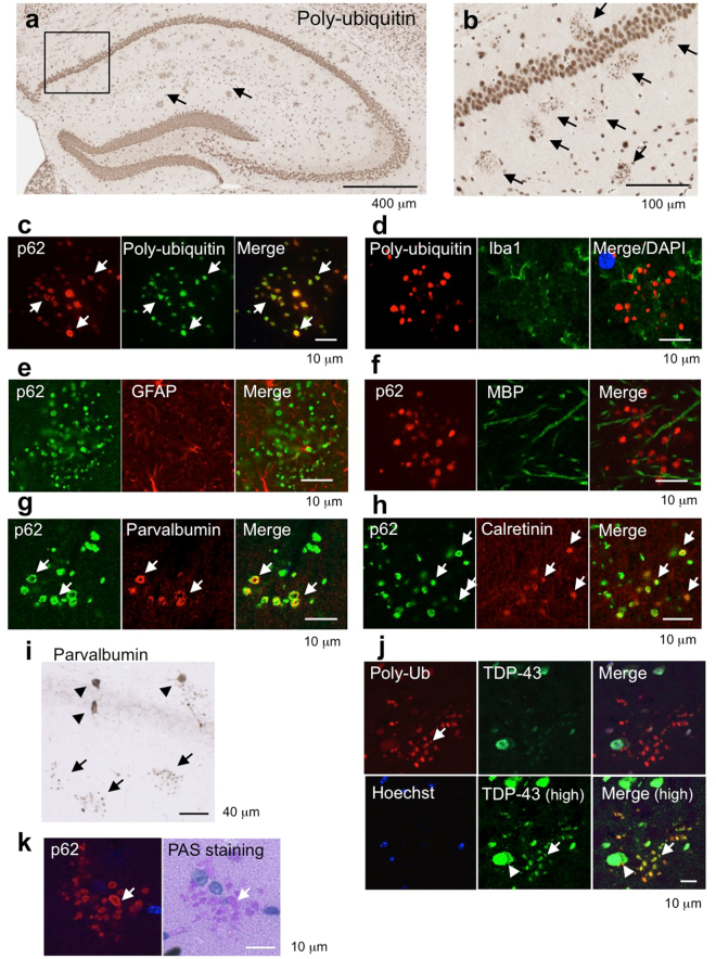
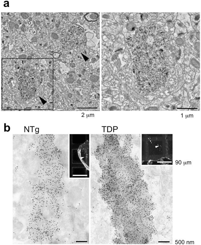
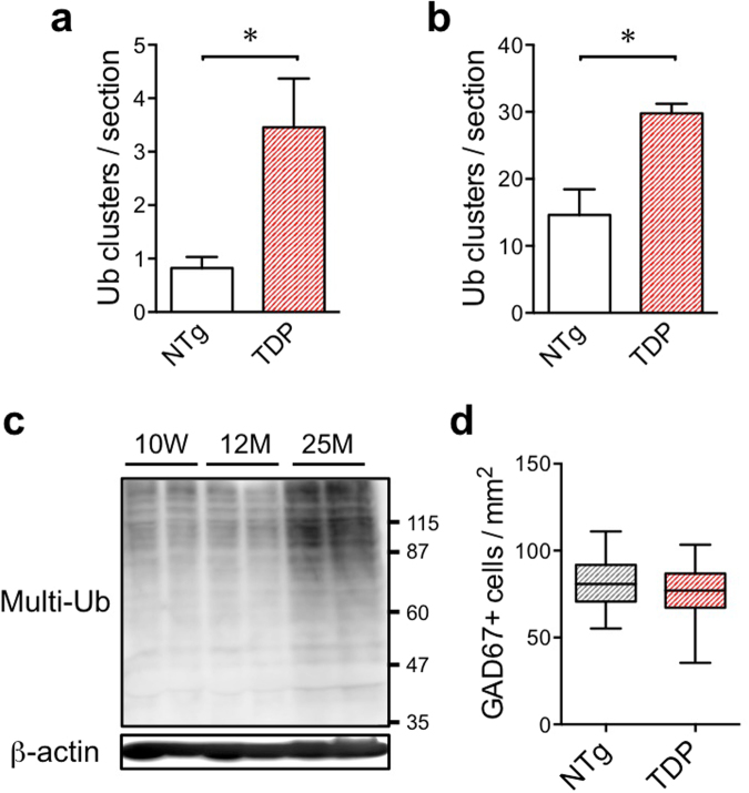
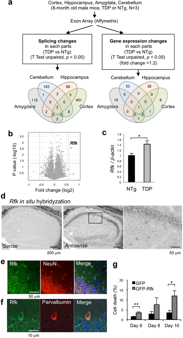
TDP-43 is an RNA-binding protein important for many aspects of RNA metabolism. Abnormal accumulation of TDP-43 in the cytoplasm of affected neurons is a pathological hallmark of the neurodegenerative diseases frontotemporal dementia (FTD) and amyotrophic lateral sclerosis (ALS). Several transgenic mouse models have been generated that recapitulate defects in TDP-43 accumulation, thus causing neurodegeneration and behavioural impairments. While aging is the key risk factor for neurodegenerative diseases, the specific effect of aging on phenotypes in TDP-43 transgenic mice has not been investigated. Here, we analyse age-dependent changes in TDP-43 transgenic mice that displayed impaired memory. We found the accumulation of abundant poly-ubiquitinated protein aggregates in the hippocampus of aged TDP-43 transgenic mice. Intriguingly, the aggregates contained some interneuron-specific proteins such as parvalbumin and calretinin, suggesting that GABAergic interneurons were degenerated in these mice. The abundance of aggregates significantly increased with age and with the overexpression of TDP-43. Gene array analyses in the hippocampus and other brain areas revealed dysregulation in genes linked to oxidative stress and neuronal function in TDP-43 transgenic mice. Our results indicate that the interneuron degeneration occurs upon aging, and TDP-43 accelerates age-dependent neuronal degeneration, which may be related to the impaired memory of TDP-43 transgenic mice.


Conflict of interest statement

The authors declare that they have no competing interests.

Figures

Figure 1
Figure 1
Generation and characterization of TDP-43 Tg mice. (a) Schematic diagram of FLAG-tagged TDP-43 and FLAG-tagged TDP-43 C-terminal fragment (R208, amino acid residue 208 to 414), which is the accumulated form of TDP-43 in the affected regions of patients with FTD/ALS, used to generate the Tg mice under the control of the mouse prion promoter. (b–d) Immunostaining of FLAG-tagged TDP-43 in TDP-43 Tg mice. The sections of the hippocampus (b), CA3 region of the hippocampus (c: the magnified image in the boxed region of Fig. 1b), and the anterior horn of the spinal cord (d) of 8-month-old TDP-43 Tg mice were stained with the anti-FLAG antibody. (e) Immunoblots of brain tissues of non-Tg littermate (NTg), heterozygous TDP-43 Tg mice (TDP) at 8 months and 18 months of age. Whole brain tissues of 8-month-old mice with the indicated genotypes were immunoblotted with anti-TDP-43, anti-FLAG, or anti-GAPDH antibodies. The filled and open arrowheads denote hTDP-43 (transgene) and mTDP-43 (endogenous), respectively. (f) Quantification of the amount of TDP-43 relative to wild-type TDP-43 in NTg mice. The intensities of bands shown in Fig. 1E were quantified using the MultiGauge software; n = 2. (g) Body weights of TDP-43 Tg mice (TDP) and non-Tg mice (NTg). Averaged body weights of the male mice with the indicated genotypes were plotted. Unpaired t-test, *p < 0.05.
Figure 2
Figure 2
Mild impairment in motor learning and severe memory deficit in heterozygous TDP-43 Tg mice. (a) Quantitative analysis of the Y-maze test data. The mean alteration rate, total number of entries, and total distance travelled by non-Tg (NTg) and TDP-43 Tg (TDP) mice are plotted. (b) Quantitative analysis of the rotarod test data. The mean holding time on the rotating rod at 8 months of age over three sequential trials is shown. (c) Experimental design used for contextual and cued fear conditioning tests. (d–g) Quantitative analysis of contextual and cued fear conditioning test data. The percentage of mean freezing times in the contextual test (d) and cued test (e) at the indicated ages is plotted. The distances travelled during conditioning of 13-month-old mice (f) or 18-month-old mice (g) are plotted for each genotype. Unpaired t-test; the data are presented as mean ± SEM. Significance level thresholds of *p < 0.05, **p < 0.01, and ***p < 0.001 were used (Fig. 3a,b,d–g).
Figure 3
Figure 3
Massive poly-ubiquitin- and p62-positive aggregates derived from GABAergic interneurons in the hippocampus of aged wild-type mice and TDP-43 Tg mice. (a,b) Immunostaining of poly-ubiquitin in the hippocampus of non-Tg mice at 20 months of age. The magnified image in the boxed region of Fig. 1a is shown (b). The arrows indicate colonies of poly-ubiquitin-positive aggregates. (ch) Immunofluorescence staining of poly-ubiquitin- and p62-positive aggregates with cell type-specific markers. The arrows indicate co-localization. The aggregates contained poly-ubiquitin and p62 (c), but not glial markers Iba1 (d), GFAP (e), or MBP (f). The aggregates contained parvalbumin (g) and calretinin (h), indicating that these are the aggregates derived from GABAergic interneurons. (i) DAB staining of the hippocampus of TDP-43 Tg mice with anti-parvalbumin (PV) antibody. The arrowheads denote intact PV neurons, while arrows denote aggregates derived from PV neurons. (j) Immunofluorescence staining of aggregates in the hippocampus of aged mice with anti-p62 and anti-TDP-43 antibodies. The arrows indicate co-localization. The arrowhead indicates cytoplasmic TDP-43. (k) Immunofluorescence staining of p62-positive aggregates following PAS staining of the hippocampus of aged mice.
Figure 4
Figure 4
Ultrastructural analyses of the cytoplasmic aggregates of the aged mice. (a) Conventional electron microscopy of the CA1 part of stratum radiatum of the hippocampus of the aged non-Tg mice. A boxed area in the left panel is enlarged and shown in the right panel. The cytoplasmic aggregates surrounded by a plasma membrane are about 1-2 µm in diameter and composed of electron-dense crystalline-like fibrillary structures. Bars: 2 µm (left) and 1 µm (right). (b) Correlative light- and immunoelectron microscopy on Tokuyasu cryosections of the hippocampus of the TDP-43 Tg (left) and aged non-Tg mice (right). 10 nm-gold particles indicating p62 were massively accumulated in the cytoplasmic aggregates corresponding to those with coarse granular fluorescence for p62 (arrows in insets). Bars: 500 nm and 90 µm (insets).
Figure 5
Figure 5
The number of aggregates derived from GABAergic interneurons are increased in TDP-43 Tg mice. (a) The averaged number of poly-ubiquitin clusters per hippocampal section of NTg and TDP-43 Tg mice at 8 months of age is plotted (mean of NTg, 0.7; mean of TDP, 2.6). The clusters bigger than 20 μm in diameter were counted. The averages of clustered number for each mouse are plotted and statistically analysed between NTg and TDP. More than 5 sections with the hippocampus bigger than 0.6 mm2 were used for each mouse. The numbers of sections used for quantification were as follows: NTg, 75 sections from 8 mice; TDP-43 Tg, 71 sections from 7 mice. Unpaired t test, *p < 0.05. (b) The averaged number of poly-ubiquitin clusters per hippocampal section of NTg and TDP-43 Tg mice at 20 months of age is plotted. The clusters bigger than 50 μm in diameter were counted. The averages of clustered number for each mouse are plotted and statistically analysed between NTg and TDP. More than 7 sections with the hippocampus bigger than 0.6 mm2 were used for each mouse. The numbers of sections used for quantification were as follows: NTg, 102 sections from 5 mice; TDP-43 Tg, 91 sections from 4 mice. Unpaired t test, *p < 0.05. (c) The increase in poly-ubiquitinated proteins in RIPA-insoluble fraction of the hippocampus of aged mice compared with those of young mice. RIPA-insoluble fractions of the hippocampus of indicated age were blotted with anti-multi-ubiquitin antibody. The same membrane was restained with anti-β-actin antibody. (d) Number of GAD67-positive neurons in the hippocampus on the section adjacent to the section used in Fig. 4j. The numbers of sections used for quantification were as follows: NTg, 44 sections from 3 mice; TDP-43 Tg, 68 sections from 3 mice.
Figure 6
Figure 6
Increased expression of riboflavin kinase in the hippocampus of TDP-43 Tg mice. (a) Experimental design of the exon array analysis. The numbers in the Venn diagram indicate the number of genes identified. (b) Plots of the expression levels of genes in the hippocampus of TDP-43 Tg mice compared with those in NTg mice. A red spot indicates the Rfk gene. (c) Quantitative PCR analysis of Rfk mRNA in the hippocampus of TDP-43 Tg mice and NTg mice. (Unpaired t-test, *p < 0.05) n = 3 each. (d) Representative images of Rfk in situ hybridisation using hippocampal sections of 8-month-old wild-type mice. Signals obtained with sense (left) and anti-sense probes (middle, right) are shown. The magnified image in the boxed region of the middle panel is shown (right). (e) Immunostaining of Rfk and NeuN in the hippocampus of 8-month-old non-Tg mice. (f) Immunostaining of Rfk and parvalbumin (PV) in the hippocampus of 8-month-old non-Tg mice. (g) Primary cultured hippocampal neurons were transfected with GFP or GFP-Rfk on day 3 of culture after dissociation of the hippocampus, and the cells were fixed and co-stained with GFP and CC3 antibodies to assess cell death. The percentage of CC3-positive cells among the total number of GFP-positive cells was calculated. The data from three independent experiments are combined and plotted. More than 200 cells for each column were counted, and more than 600 cells were counted for day 6. The data are presented as mean ± SEM. Unpaired t-test, *p < 0.05, **p < 0.01.

References

    1. Renton AE, Chio A, Traynor BJ. State of play in amyotrophic lateral sclerosis genetics. Nat Neurosci. 2014;17:17–23. doi: 10.1038/nn.3584. - DOI - PMC - PubMed
    1. Neumann M, et al. Ubiquitinated TDP-43 in frontotemporal lobar degeneration and amyotrophic lateral sclerosis. Science. 2006;314:130–133. doi: 10.1126/science.1134108. - DOI - PubMed
    1. Arai T, et al. TDP-43 is a component of ubiquitin-positive tau-negative inclusions in frontotemporal lobar degeneration and amyotrophic lateral sclerosis. Biochem Biophys Res Commun. 2006;351:602–611. doi: 10.1016/j.bbrc.2006.10.093. - DOI - PubMed
    1. Ling SC, Polymenidou M, Cleveland DW. Converging mechanisms in ALS and FTD: disrupted RNA and protein homeostasis. Neuron. 2013;79:416–438. doi: 10.1016/j.neuron.2013.07.033. - DOI - PMC - PubMed
    1. Li YR, King OD, Shorter J, Gitler AD. Stress granules as crucibles of ALS pathogenesis. J Cell Biol. 2013;201:361–372. doi: 10.1083/jcb.201302044. - DOI - PMC - PubMed

Publication types

MeSH terms