Review
. 2017 Mar 20:2017:7064120.
doi: 10.1155/2017/7064120. eCollection 2017.

Clinical Applications of Contrast-Enhanced Perfusion MRI Techniques in Gliomas: Recent Advances and Current Challenges

Affiliations
Review

Clinical Applications of Contrast-Enhanced Perfusion MRI Techniques in Gliomas: Recent Advances and Current Challenges

Junfeng Zhang et al. Contrast Media Mol Imaging. .

Abstract

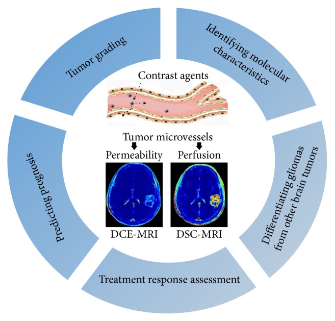
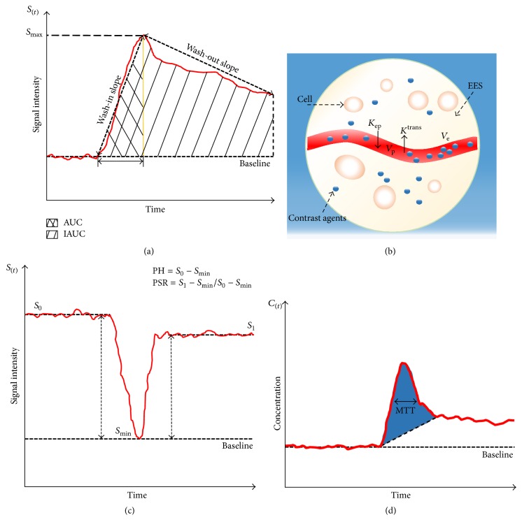
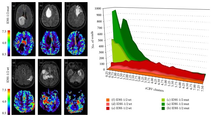
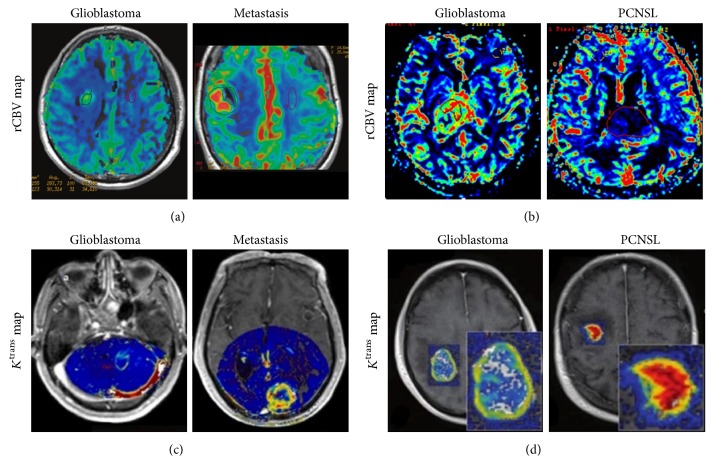
Gliomas possess complex and heterogeneous vasculatures with abnormal hemodynamics. Despite considerable advances in diagnostic and therapeutic techniques for improving tumor management and patient care in recent years, the prognosis of malignant gliomas remains dismal. Perfusion-weighted magnetic resonance imaging techniques that could noninvasively provide superior information on vascular functionality have attracted much attention for evaluating brain tumors. However, nonconsensus imaging protocols and postprocessing analysis among different institutions impede their integration into standard-of-care imaging in clinic. And there have been very few studies providing a comprehensive evidence-based and systematic summary. This review first outlines the status of glioma theranostics and tumor-associated vascular pathology and then presents an overview of the principles of dynamic contrast-enhanced MRI (DCE-MRI) and dynamic susceptibility contrast-MRI (DSC-MRI), with emphasis on their recent clinical applications in gliomas including tumor grading, identification of molecular characteristics, differentiation of glioma from other brain tumors, treatment response assessment, and predicting prognosis. Current challenges and future perspectives are also highlighted.


Figures

Figure 1
Figure 1
The versatile clinical applications of contrast-enhanced perfusion MRI techniques in gliomas.
Figure 2
Figure 2
An illustration of parameters derived from DCE-MRI and DSC-MRI. (a) Semiquantitative parameters from signal intensity curve in DCE-MRI. (b) Schematic diagram of ETK model from DCE-MRI. (c) Calculation of PSR and PH from DSC-MRI. (d) Contrast concentration-time course curve of DSC-MRI. CBV is proportional to determined area under contrast concentration-time course curve (blue shaded area), and CBF is easily calculated given the relationship of MTT and CBV.
Figure 3
Figure 3
DSC-MRI for identification of IDH mutation status in GBM. Six sets of representative FLAIR and corresponding rCBV images from IDH1/2 mutant and wild-type GBM. Histogram analysis demonstrates that IDH1/2 mutant tumors have substantially lower rCBV value than the wild-type. Reproduce with permission from Kickingereder et al. [60].
Figure 4
Figure 4
DSC-MRI (a) and DCE-MRI (b) for differentiation of GBM, PCNSL, and metastasis. rCBV maps demonstrate different characteristic features in the three distinct entities, with significantly higher rCBV value of GBM compared with metastasis and PCNSL. The Ktrans value of GBM is significantly lower than metastasis and PCNSL. Reproduce with permission from Mangla et al. [118], Xing et al. [120], Zhao et al. [52], and Kickingereder et al. [121].
Figure 5
Figure 5
Discrimination of PsP from PD using DSC-MRI and DCE-MRI. (a) Contrast-enhanced T1WI of GBM treated with temozolomide demonstrates increased contrast enhancement suspicious for both PsP (top row) and PD (bottom row). Corresponding rCBV maps show low perfusion in PsP and high perfusion in PD; (b) Ktrans maps demonstrate decreased Ktrans value in PsP (top row) compared with PD (bottom row). Reproduce with permission from Shin et al. [158] and Thomas et al. [161].
Figure 6
Figure 6
Discrimination of RN from recurrent GBM using DCE-MRI (a) and DSC-MRI (b). Contrast-enhanced T1WI demonstrates similar contrast enhancement in recurrent glioblastoma (top row) and RN (bottom row). Corresponding rCBV and Ktrans maps show significant difference between these two entities, with higher Ktrans and rCBV for recurrent tumor (top row) but low for RN (bottom row). Reproduce with permission from Bisdas et al. [184] and Masch et al. [186].

References

    1. Louis D. N., Perry A., Reifenberger G., et al. The 2016 World Health Organization Classification of Tumors of the Central Nervous System: a summary. Acta Neuropathologica. 2016;131(6):803–820. doi: 10.1007/s00401-016-1545-1. - DOI - PubMed
    1. Wen P. Y., Reardon D. A. Neuro-oncology in 2015: progress in glioma diagnosis, classification and treatment. Nature Reviews Neurology. 2016;12(2):69–70. doi: 10.1038/nrneurol.2015.242. - DOI - PubMed
    1. Weller M., van den Bent M., Hopkins K., et al. EANO guideline for the diagnosis and treatment of anaplastic gliomas and glioblastoma. The Lancet Oncology. 2014;15(9):e395–e403. doi: 10.1016/S1470-2045(14)70011-7. - DOI - PubMed
    1. Hardee M. E., Zagzag D. Mechanisms of glioma-associated neovascularization. American Journal of Pathology. 2012;181(4):1126–1141. doi: 10.1016/j.ajpath.2012.06.030. - DOI - PMC - PubMed
    1. Kimura M., da Cruz L. C. Multiparametric MR imaging in the assessment of brain tumors. Magnetic Resonance Imaging Clinics of North America. 2016;24(1):87–122. doi: 10.1016/j.mric.2015.09.001. - DOI - PubMed

Publication types