. 2017 Aug 8:2017:9481276.
doi: 10.1155/2017/9481276. eCollection 2017.

Correlation of Somatostatin Receptor-2 Expression with Gallium-68-DOTA-TATE Uptake in Neuroblastoma Xenograft Models

Affiliations

Correlation of Somatostatin Receptor-2 Expression with Gallium-68-DOTA-TATE Uptake in Neuroblastoma Xenograft Models

Libo Zhang et al. Contrast Media Mol Imaging. .

Abstract

Peptide-receptor imaging and therapy with radiolabeled somatostatin analogs such as 68Ga-DOTA-TATE and 177Lu-DOTA-TATE have become an effective treatment option for SSTR-positive neuroendocrine tumors. The purpose of this study was to evaluate the correlation of somatostatin receptor-2 (SSTR2) expression with 68Ga-DOTA-TATE uptake and 177Lu-DOTA-TATE therapy in neuroblastoma (NB) xenograft models. We demonstrated variable SSTR2 expression profiles in eight NB cell lines. From micro-PET imaging and autoradiography, a higher uptake of 68Ga-DOTA-TATE was observed in SSTR2 high-expressing NB xenografts (CHLA-15) compared to SSTR2 low-expressing NB xenografts (SK-N-BE(2)). Combined autoradiography-immunohistochemistry revealed histological colocalization of SSTR2 and 68Ga-DOTA-TATE uptake in CHLA-15 tumors. With a low dose of 177Lu-DOTA-TATE (20 MBq/animal), tumor growth inhibition was achieved in the CHLA-15 high SSTR2 expressing xenograft model. Although, in vitro, NB cells showed variable expression levels of norepinephrine transporter (NET), a molecular target for 131I-MIBG therapy, low 123I-MIBG uptake was observed in all selected NB xenografts. In conclusion, SSTR2 expression levels are associated with 68Ga-DOTA-TATE uptake and antitumor efficacy of 177Lu-DOTA-TATE. 68Ga-DOTA-TATE PET is superior to 123I-MIBG SPECT imaging in detecting NB tumors in our model. Radiolabeled DOTA-TATE can be used as an agent for NB tumor imaging to potentially discriminate tumors eligible for 177Lu-DOTA-TATE therapy.


Figures

Figure 1
Figure 1
mRNA expression level and Western blotting analyses of SSTR2 and NET. (a) RNA was isolated from different neuroblastoma cell lines, converted into cDNA, followed by RT-PCR with SSTR2 and NET specific primers. The GAPDH gene was used as a reference gene. (b) Protein lysates were prepared from different neuroblastoma cell lines. Protein samples were separated by polyacrylamide gel electrophoresis. Expression of SSTR2 and NET proteins was visualized using specific antibodies. β-Actin was used as internal loading control.
Figure 2
Figure 2
Representative micro-PET/CT images at 1 hour after injection of 10 MBq of  68Ga-DOTA-TATE in the CHLA-15 (a) and SK-N-BE(2) (b) tumor-bearing NOD/SCID mice. Images were presented in the axial (left) and coronal (right) orientations. The white arrows denote localized tumor on the shoulder. (c) Standardized Uptake Values (SUV) in CHLA-15 and SK-N-BE(2) xenografts were calculated using the formula: SUV = CPET(T)/(Injected  dose/Bodyweight). The difference was significant between CHLA-5 and SK-N-BE(2) tumors (∗∗P < 0.01). Two-tailed unpaired t-tests were performed to compare the SUVmean values obtained.
Figure 3
Figure 3
Colocalization of SSTR2 and autoradiography on CHLA-15 xenografts. CHLA-15 and SK-N-BE(2) tumors were removed immediately after PET/CT imaging. The spatial distribution of the 68Ga-DOTA-TATE uptake was visualized by autoradiography (white signal) in the serial sections of CHLA-15 (a) and SK-N-BE(2) (b) tumors. (c) Representative CHLA-15 tumor section was stained for SSTR2 (red fluorescence) and DAPI (blue fluorescence). (d) The merging image of SSTR2 immunostaining and autoradiograph of the same CHLA-15 tumor section. (e) A representative SK-N-BE(2) tumor section stained for SSTR2 (red fluorescence) and DAPI (blue fluorescence).
Figure 4
Figure 4
Antitumoral effects of 177Lu-DOTA-TATE in the CHLA-15 neuroblastoma model. (a) Mice (n = 7) with subcutaneous CHLA-15 xenografts were treated with one dose of 20 MBq of 177Lu-DOTA-TATE. Control mice (n = 7) received saline. Tumor volume was measured and plotted as shown. Values are stated as mean ± SE; P < 0.05. (b) Animal body weight was monitored in CHLA-15 tumor-bearing mice with/without 177Lu-DOTA-TATE treatment.
Figure 5
Figure 5
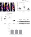
(a) Example images of 123I-MIBG SPECT/CT in the SK-N-BE(2), CHLA-15, and BE(2)C xenograft models. The white arrows denote localized tumor on the shoulder. (b) Scatter plots of the tumor-to-muscle (T/M) ratios of 123I-MIBG uptake in the SK-N-BE(2), CHLA-15, and BE(2)C xenograft models derived from SPECT/CT imaging. One-way analysis of variance (ANOVA) with Tukey's test was used for statistical analysis. P<0.05; ∗∗P < 0.01. (c) Scatter plots of the tumor-to-muscle (T/M) ratio of 123I-MIBG autoradiography in the SK-N-BE(2), CHLA-15, and BE(2)C xenograft models. One-way ANOVA with Tukey's test was used for statistical analysis. P < 0.05; ∗∗P<0.01. (d) Western Blot of NET with homogenized CHLA-15, SK-N-BE(2), and BE(2)C xenografts. Three tumors were randomly selected from three groups for Western blot analysis. (e) Quantitative analysis of NET protein expression in CHLA-15, SK-N-BE(2), and BE(2)C xenografts. NET protein expression in Western blot images was quantified densitometrically using ImageJ software (NIH, USA) and normalized with respect to the corresponding expression of β-actin. Comparison of NET expression between different groups was analyzed by the nonparametric Kruskal-Wallis analysis with Dunn's multiple comparison tests. No significant difference of NET expression was observed between CHLA-15, SK-N-BE(2), and BE(2)C xenografts.

References

    1. Howman-Giles R., Shaw P. J., Uren R. F., Chung D. K. V. Neuroblastoma and Other Neuroendocrine Tumors. Seminars in Nuclear Medicine. 2007;37(4):286–302. doi: 10.1053/j.semnuclmed.2007.02.009. - DOI - PubMed
    1. Mugishima H. Current status of molecular biology and treatment strategy for neuroblastoma. International Journal of Clinical Oncology. 2012;17(3):p. 189. doi: 10.1007/s10147-012-0423-7. - DOI - PubMed
    1. Ora I., Eggert A. Progress in treatment and risk stratification of neuroblastoma: impact on future clinical and basic research. Seminars in Cancer Biology. 2011;21(4):217–228. doi: 10.1016/j.semcancer.2011.07.002. - DOI - PubMed
    1. Maecke H. R., Reubi J. C. Somatostatin receptors as targets for nuclear medicine imaging and radionuclide treatment. Journal of Nuclear Medicine. 2011;52(6):841–844. doi: 10.2967/jnumed.110.084236. - DOI - PubMed
    1. Georgantzi K., Tsolakis A. V., Stridsberg M., Jakobson Å., Christofferson R., Janson E. T. Differentiated expression of somatostatin receptor subtypes in experimental models and clinical neuroblastoma. Pediatric Blood and Cancer. 2011;56(4):584–589. doi: 10.1002/pbc.22913. - DOI - PubMed

LinkOut - more resources