. 2017 Nov;14(5):5484-5490.
doi: 10.3892/ol.2017.6846. Epub 2017 Aug 28.

Role of cytochrome P450 2J2 on cell proliferation and resistance to an anticancer agent in hepatocellular carcinoma HepG2 cells

Affiliations

Role of cytochrome P450 2J2 on cell proliferation and resistance to an anticancer agent in hepatocellular carcinoma HepG2 cells

Geun Hye Hwang et al. Oncol Lett. 2017 Nov.

Abstract

The present study examined the role of human cytochrome P450 2J2 (CYP2J2) on cell proliferation and resistance to an anticancer agent using stable hepatocellular carcinoma HepG2 cells overexpressing CYP2J2. Overexpression of CYP2J2 significantly increased HepG2 cell proliferation and the expression levels of cell cycle regulatory proteins, including cyclin D1, cyclin E, cyclin-dependent kinase (Cdk)2 and Cdk4. CYP2J2-overexpressing HepG2 cells exhibited high levels of Akt phosphorylation compared with those observed in wild-type HepG2 cells. Although Akt phosphorylation in both cell lines was significantly attenuated by LY294002, a specific phosphoinositide 3-kinase/Akt signaling inhibitor, the levels of Akt phosphorylation following treatment with LY294002 were higher in CYP2J2-overexpressing HepG2 cells than in wild-type HepG2 cells. Cell counting revealed that proliferation was reduced by LY294002 in both cell lines; however, CYP2J2-overexpressing HepG2 cell numbers were higher than those of wild-type HepG2 cells following treatment with LY294002. These results indicated that increased cell proliferation by CYP2J2 overexpression is mediated by increased Akt activity. It was also demonstrated that doxorubicin, an anticancer agent, reduced cell viability, induced a significant increase in the B-cell lymphoma (Bcl)-2 associated X protein (Bax)/Bcl-2 ratio and decreased pro-caspase-3 levels in wild-type HepG2 cells. However, the doxorubicin-induced reduction in cell viability was significantly attenuated by enhanced upregulation of CYP2J2 expression. The increase in the Bax/Bcl-2 ratio and the decrease in pro-caspase-3 levels were also recovered by CYP2J2 overexpression. In conclusion, CYP2J2 serves important roles in cancer cell proliferation and resistance to the anticancer agent doxorubicin in HepG2 cells.

Keywords: CYP2J2; HepG2 cells; cancer; proliferation; resistance.


Figures
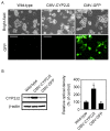
Figure 1.
Figure 1.
Effect of overexpression of CYP2J2 in HepG2 cells. (A) Representative images of wild-type, CMV-CYP2J2 and CMV-GFP-transfected HepG2 cells. Magnification, ×200. Scale bar, 50 µm. (B) Cells were infected with either CMV-CYP2J2 or CMV-GFP lentiviruses, and cell lysates were subjected to western blot analysis using an anti-CYP2J2-specific antibody. Charts represent three experiments. Values are expressed relative to the β-actin levels. Error bars denote the mean ± SEM for three independent experiments. *P<0.05 vs. wild-type. CYP2J2, cytochrome P450 2J2; SEM, standard error of the mean; GFP, green fluorescent protein; CMV, cytomegalovirus.
Figure 2.
Figure 2.
Effect of overexpression of CYP2J2 on cell proliferation. (A) Cell proliferation rates in wild-type and CMV-CYP2J2-transfected HepG2 cells were assessed by direct cell counting at the indicated time points. Error bars denote the mean ± SEM for three independent experiments levels. *P<0.05 vs. control. (B) The levels of cyclin D1, cyclin E, Cdk4 and Cdk2 in wild-type, CMV-CYP2J2- and CMV-GFP-transfected cells were assessed by western blot analysis. Each of the examples shown is representative of three experiments. The graphs denote the mean ± SEM of three independent experiments for each condition, as determined from densitometry analysis relative to β-actin. *P<0.05 vs. wild-type. CYP2J2, cytochrome P450 2J2; SEM, standard error of the mean; GFP, green fluorescent protein; CMV, cytomegalovirus; Cdk, cyclin-dependent kinase.
Figure 3.
Figure 3.
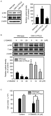
Involvement of the Akt signaling pathway in CYP2J2-induced cell proliferation. (A) The expression levels of phosphorylation of Akt (Ser473) in wild-type, CMV-CYP2J2- and CMV-GFP-transfected HepG2 cells were assessed by western blot analysis. Each of the examples shown is representative of three experiments. The graphs denote the mean ± SEM of three independent experiments for each condition, as determined from densitometry analysis relative to β-actin. *P<0.05 vs. wild-type. (B) Wild-type and CMV-CYP2J2-transfected HepG2 cells were treated with LY294002 (0–20 µM) for 8 h, and the phosphorylation of Akt was then detected by western blot analysis. Each of the examples shown is representative of three experiments. The graphs denote the mean ± SEM of three independent experiments for each condition, as determined from densitometry analysis relative to β-actin. *P<0.05 vs. wild-type. (C) Wild-type and CMV-CYP2J2-transfected HepG2 cells were cultured for 48 h, and the cells were then cultured with or without 10 µM LY294002 for additional 24 h. Cell proliferation rates in wild-type and CMV-CYP2J2-transfected cells were assessed by direct cell counting. Error bars denote the mean ± SEM of three independent experiments levels. *P<0.05 vs. control; #P<0.05 vs. wild-type. CYP2J2, cytochrome P450 2J2; SEM, standard error of the mean; GFP, green fluorescent protein; CMV, cytomegalovirus; Cdk, cyclin-dependent kinase; p, phosphorylated; T, total.
Figure 4.
Figure 4.
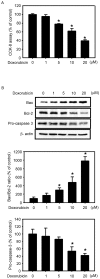
Effect of doxorubicin on HepG2 cells. (A) HepG2 cells were treated with the indicated concentration of doxorubicin for 24 h, and cell viability was measured using a CCK-8 reduction assay. Values are expressed as the mean ± SEM of three experiments with triplicate dishes. *P<0.05 vs. control. (B) HepG2 cells were cultured under the indicated conditions for 24 h. Cell lysates were subjected to western blot analysis using anti-Bax, anti-Bcl-2 and anti-pro-caspase-3-specific antibodies. Each of the examples shown is representative of three experiments. The intensities of Bax, Bcl-2 and pro-caspase-3 bands were determined by densitometry analysis relative to β-actin, and the Bax/Bcl-2 ratio was calculated. Values are expressed as the mean ± SEM of three independent experiments. *P<0.05 vs. control. CCK, Cell Counting kit; Bcl, B-cell lymphoma; Bax, Bcl-2 associated X protein; SEM, standard error of the mean.
Figure 5.
Figure 5.
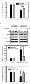
Effect of CYP2J2 overexpression on the resistance to doxorubicin. (A) Wild-type and CMV-CYP2J2-transfected HepG2 cells were cultured with or without 10 µM doxorubicin for 24 h, and cell viability was measured using a CCK-8 reduction assay. Values are expressed as the mean ± SEM of three experiments with triplicate dishes. *P<0.05 vs. control; #P<0.05 vs. wild-type. (B) Wild-type and CMV-CYP2J2-transfected HepG2 cells were cultured under the indicated conditions for 24 h. Cell lysates were subjected to western blot analysis using anti-Bax, anti-Bcl-2 and anti-pro-caspase-3-specific antibodies. Each of the examples shown is representative of three experiments. The intensities of the Bax, Bcl-2 and pro-caspase-3 bands were determined by densitometry analysis relative to β-actin, and the Bax/Bcl-2 ratio was calculated. Values are expressed as the mean ± SEM of three independent experiments. *P<0.05 vs. control; #P<0.05 vs. wild-type. CCK, Cell Counting kit; Bcl, B-cell lymphoma; Bax, Bcl-2 associated X protein; SEM, standard error of the mean; CYP2J2, cytochrome P450 2J2; SEM, standard error of the mean; CMV, cytomegalovirus.

References

    1. Capdevila JH, Falck JR, Harris RC. Cytochrome P450 and arachidonic acid bioactivation. Molecular and functional properties of the arachidonate monooxygenase. J Lipid Res. 2000;41:163–181. - PubMed
    1. Enayetallah AE, French RA, Thibodeau MS, Grant DF. Distribution of soluble epoxide hydrolase and of cytochrome P450 2C8, 2C9 and 2J2 in human tissues. J Histochem Cytochem. 2004;52:447–454. doi: 10.1177/002215540405200403. - DOI - PubMed
    1. Fisslthaler B, Popp R, Kiss L, Potente M, Harder DR, Fleming I, Busse R. Cytochrome P450 2C is an EDHF synthase in coronary arteries. Nature. 1999;401:493–497. doi: 10.1038/46816. - DOI - PubMed
    1. Rosolowsky M, Campbell WB. Synthesis of hydroxyeicosatetraenoic (HETEs) and epoxyeicosatrienoic acids (EETs) by cultured bovine coronary artery endothelial cells. Biochim Biophys Acta. 1996;1299:267–277. doi: 10.1016/0005-2760(95)00216-2. - DOI - PubMed
    1. Lee HC, Lu T, Weintraub NL, VanRollins M, Spector AA, Shibata EF. Effects of epoxyeicosatrienoic acids on the cardiac sodium channels in isolated rat ventricular myocytes. J Physiol. 1999;1:153–168. doi: 10.1111/j.1469-7793.1999.0153o.x. - DOI - PMC - PubMed