. 2017 Dec 19;16(1):62-69.
doi: 10.1039/c7ob02453c.

Synthesis and biological assessment of 3,7-dihydroxytropolones

Affiliations

Synthesis and biological assessment of 3,7-dihydroxytropolones

D R Hirsch et al. Org Biomol Chem. .

Abstract

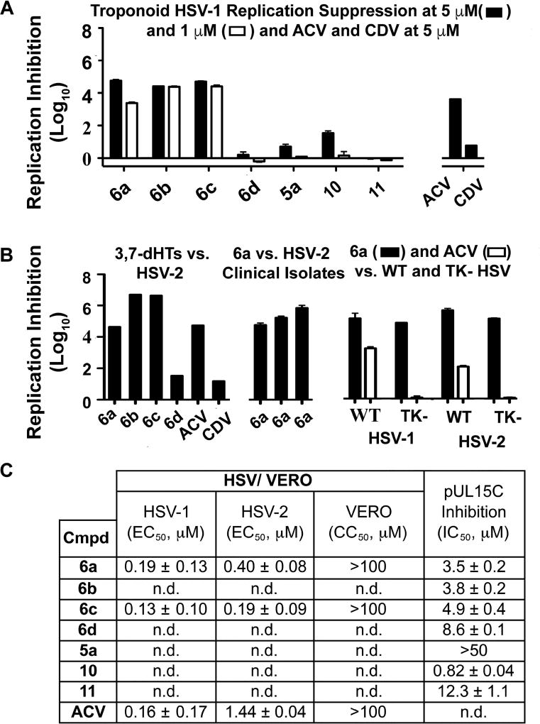
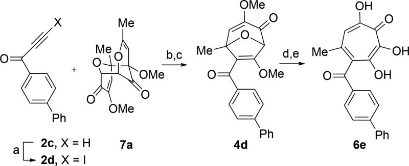
3,7-Dihydroxytropolones (3,7-dHTs) are highly oxygenated troponoids that have been identified as lead compounds for several human diseases. To date, structure-function studies on these molecules have been limited due to a scarcity of synthetic methods for their preparation. New synthetic strategies towards structurally novel 3,7-dHTs would be valuable in further studying their therapeutic potential. Here we describe the successful adaptation of a [5 + 2] oxidopyrilium cycloaddition/ring-opening for 3,7-dHT synthesis, which we apply in the synthesis of a plausible biosynthetic intermediate to the natural products puberulic and puberulonic acid. We have also tested these new compounds in several biological assays related to human immunodeficiency virus (HIV), hepatitis B virus (HBV) and herpes simplex virus (HSV) in order to gain insight into structure-functional analysis related to antiviral troponoid development.


Conflict of interest statement

Conflict of Interest

RM, LM, and JT are coinventors on a patent application that covers anti-HBV and anti-HSV activity of hydroxytropolones.

Figures

Figure 1
Figure 1
Viral ribonuclease H-related studies. (A) Compounds tested in antiviral assays. (B) Summary of data from RNase H-related biological studies for HIV and HBV. a HIV-1 RT RNaseH IC50 values are reported as the average from triplicate assays ± standard deviation.b Concentration of 50% viral replication suppression (EC50) are reported from a single run. n.p. Reflects not protective. c CC50 represents concentration at 50% cytotoxicity against the host CEM-SS cells, and are calculated from a single experiment. d HBV replication inhibition at 20 µM, unless otherwise defined. ** Indicates that the amount of (+)-strand DNA is <25% and (−)-strand DNA is >60% relative to DMSO control. * Indicates that the amount of (+)-strand DNA is <50% of the amount of (−)-strand DNA relative to DMSO control. – refers to no evidence of selective inhibition of (+)-strand DNA. EC50 values are shown for active compounds, and are reported as the average of two or three dose response curves ± standard deviation. e HepDES19 cytotoxicity values are likewise reported as an average of 2–4 assays ± standard deviation.
Figure 2
Figure 2
HSV-related studies. (A) HSV-1 suppression by synthetic tropolones at concentrations of 5 and 1 µM. Also shown are clinical anti-HSV agents acyclovir (ACV) and cidofovir (CDV) at 5 µM. (B) Replication inhibition studies of 3,7-dHTs at 5 µM. L/R 6a–d against HSV-2, compound 6a against 3 HSV-2 clinical isolates, and comparisons of 6a and ACV against wild-type (WT) and thymidine kinase negative (TK) HSV strains resistant to ACV. (C) EC50 values obtained for 6a and 6c against HSV-1 and HSV-2, with data representing the average from 2 dose response curves +/− standard deviation. Also shown are values for ACV obtained previously by Tavis et al under identical conditions. CC50 represents concentration at 50% cytotoxicity against the host Vero cells. n.d. = not determined.
Figure 3
Figure 3
HSV-Associated Assays of Compound 12 and Related Structures. (A) Structures of most potent anti-HSV molecules. (B) Head-to-head comparison of troponoids as HSV-1 viral replication inhibitors at three concentrations. (C) Potency values for 6e and 12. Values of 12 were reported previously.
Figure 4
Figure 4
Hypothesized Structural Basis for Potency Increases of 6e against pUL15. (A) Single orientation of αHT as opposed to (B) two orientations of 3,7-dHTs. (C,D) The structure of the complex between UL15 (PDB id 4IOX) and 3,7-dHT 6e (green) predicted by molecular docking, showing (C) the biphenyl group of the ligand nested into a hydrophobic groove formed by Lys 640, Asn 583 and Leu 636 and (D) possible π-cation contacts with Lys 640 and close proximity to Asn 583, which could participate in π-NH interactions.
Scheme 1
Scheme 1
(A) Contiguously hydroxylated tropolones and common mode of metalloenzyme binding. (B) A divergent, unifying galactose approach towards both tropolone classes. (C) Overview of kojic acid route developed by our lab and extended in this study to 3,7-dHTs.
Scheme 2
Scheme 2
(a) Formation of 3,7-dHTs via oxidopyrylium salt 1a and iodoalkynes 2a and 2b. (b) Results from initial attempts at methanol-iodide exchange. (c) Brønsted acid-mediated cycloreversion of oxabicyclic intermediates 3. (d) Acid-mediated hydrolysis and decarboxylation of 5a. Reagents and conditions: (a) i-Pr2NPh, 2a/b, CH2Cl2, 120 °C, 20 min (3a = 57%, 3b = 95%). (b) MeOH/DMAP, 120 °C, 15 min (4a = 94%, 4b = 73%). (c) BCl3, CH2Cl2, r.t., 10 min, (5a = 97%,5b = 81%). (d) HBr/AcOH, 120 °C, 45 min (6a = 55%, 6b = 87%). (e) DMAP/H2O, CDCl3, 100 °C, 90 min, 16%. (f) K2CO3/MeOH, r.t., 67%. (g) HBr/AcOH/H2O, 120 °C, 60 min, 45%.
Scheme 3
Scheme 3
Biogenesis of 6-hydroxytropolones stipitatonic acid and stipitatic acid from stipitalide and related 3,7-dHTs.
Scheme 4
Scheme 4
Synthesis of 3,7-dHT 6d. Reagents and conditions: (a) iPr2NPh, 2b, CH2Cl2, 100 °C, 1 hr, 5:1 ratio of 3c:3b observed by crude 1H NMR. (b) NEt3, CH2Cl2, 85%. (c) 2b, CH2Cl2, 100 °C, 1 hr, 71%. (d) DMAP, MeOH, 120 °C, 5 min, 61%. (e) BCl3, CH2Cl2, 0 °C, 5 min, 89%. (f) NaOAc, AcOH, r.t., 15 hr, 93%. (g) NaOH (aq.), r.t., 3 hr, 91%. (h) C18 silica, MeCN/H2O/TFA, 81%. (i) HBr/AcOH, 120 °C, 45 min, 35%.
Scheme 5
Scheme 5
Synthesis of 3,7-dHT 6e. Reagents and conditions: (a) AgNO3, N-iodosuccinimide, acetone, 0°C, 4 hr, 57%. (b) 2d and 7a, CH2Cl2, 120°C, 30 min. (c) MeOH/DMAP, 70°C, 15 min, 68% over two steps. (d) BCl3, CH2Cl2, 0 °C, 6 min, 64%. (e) HBr/AcOH, 120°C, 70 min, 70%.

References

    1. For a review, see: Meck C, D’Erasmo MP, Hirsch DR, Murelli RP. Med. Chem. Commun. 2014;5:842–852.

    1. Didierjean J, Isel C, Querré F, Mouscadet JF, Aubertin AM, Valnot JV, Piettre SR, Marquet R. Antimicrob. Agents Chemother. 2005;49:4884–4894. - PMC - PubMed
    1. Budihas SR, Gorshkova I, Gaidamakov S, Wamiru A, Bona MK, Parniak MMA, Crouch RJ, McMahon JB, Beutler JA, Le Grice SFJ. Nucleic Acids Res. 2005;33:1249–1256. - PMC - PubMed
    1. Allen NE, Alborn WE, Jr, Hobbs JN, Jr, Kirst HA. Antimicrob Agents Chemother. 1982;22:824. - PMC - PubMed
    1. Iwatsuki M, Takada S, Mori M, Ishiyama A, Namatame M, Otoguro K, Nishihara-Tsukashima A, Nonaka K, Masuma R, Otoguro O, Shiomi K, Omura S. J. Antibiot. 2011;64:183–188. - PubMed

MeSH terms