. 2019 Apr;269(4):756-766.
doi: 10.1097/SLA.0000000000002504.

Electric Field Based Dressing Disrupts Mixed-Species Bacterial Biofilm Infection and Restores Functional Wound Healing

Affiliations

Electric Field Based Dressing Disrupts Mixed-Species Bacterial Biofilm Infection and Restores Functional Wound Healing

Kasturi Ganesh Barki et al. Ann Surg. 2019 Apr.

Abstract

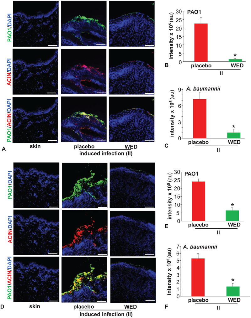
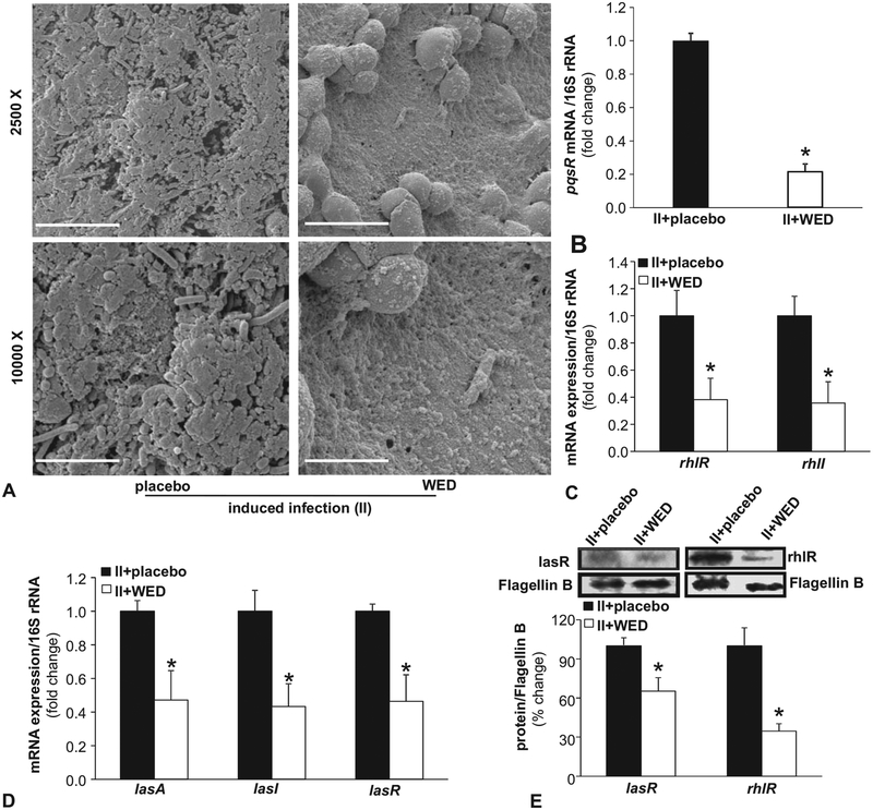
Objective: This study was designed to employ electroceutical principles, as an alternative to pharmacological intervention, to manage wound biofilm infection. Mechanism of action of a United States Food and Drug Administration-cleared wireless electroceutical dressing (WED) was tested in an established porcine chronic wound polymicrobial biofilm infection model involving inoculation with Pseudomonas aeruginosa PAO1 and Acinetobacter baumannii 19606.

Background: Bacterial biofilms represent a major wound complication. Resistance of biofilm toward pharmacologic interventions calls for alternative therapeutic strategies. Weak electric field has anti-biofilm properties. We have previously reported the development of WED involving patterned deposition of Ag and Zn on fabric. When moistened, WED generates a weak electric field without any external power supply and can be used as any other disposable dressing.

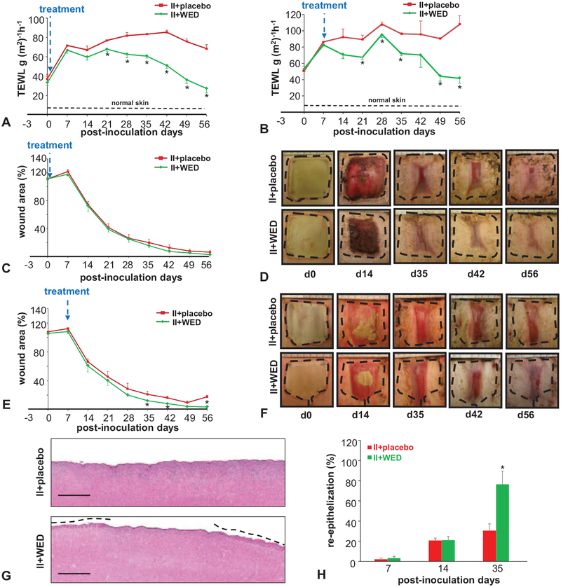
Methods: WED dressing was applied within 2 hours of wound infection to test its ability to prevent biofilm formation. Alternatively, WED was applied after 7 days of infection to study disruption of established biofilm. Wounds were treated with placebo dressing or WED twice a week for 56 days.

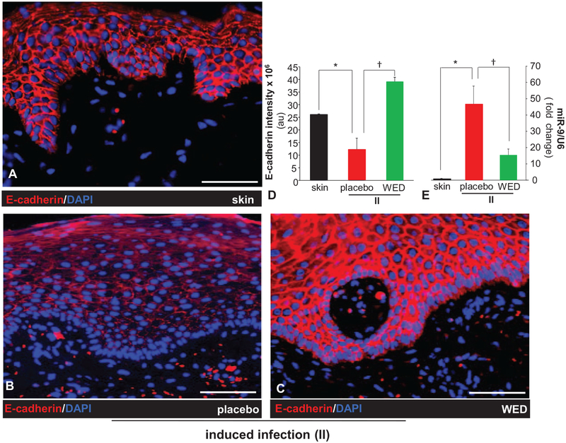
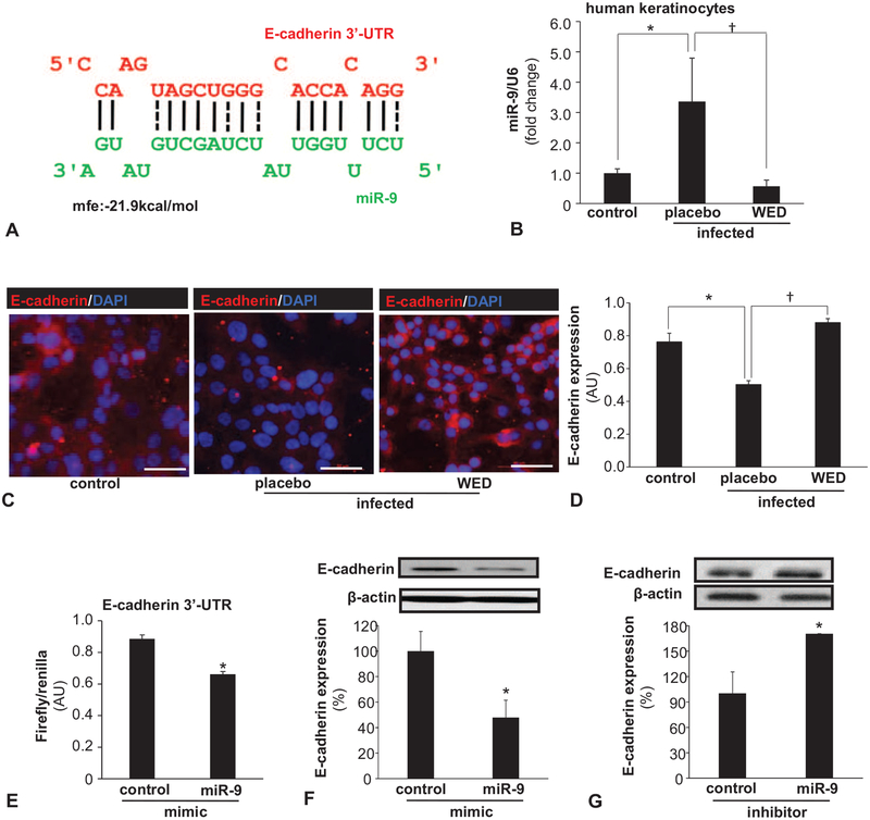
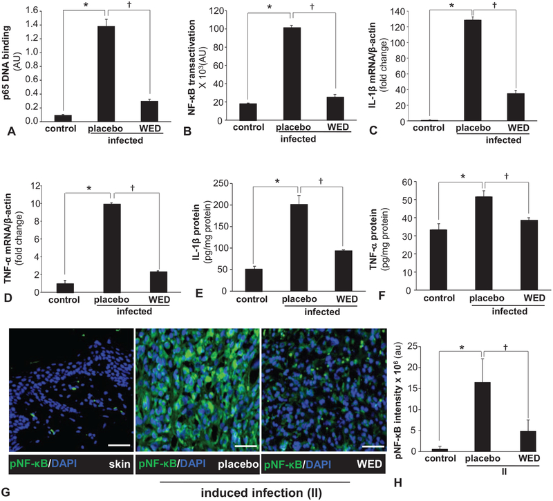
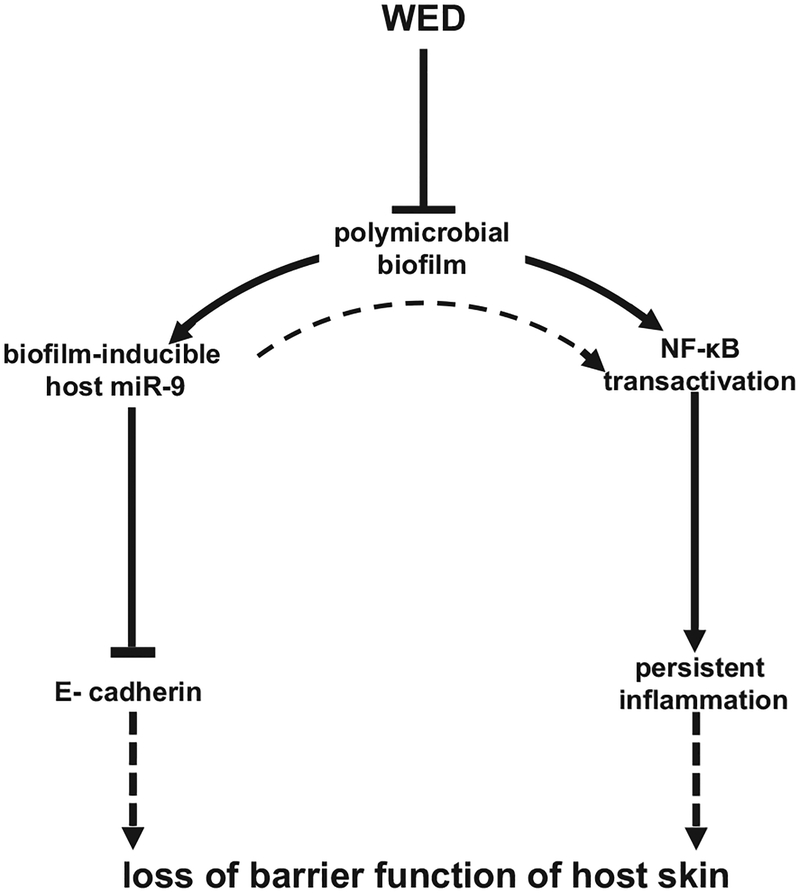
Results: Scanning electron microscopy demonstrated that WED prevented and disrupted wound biofilm aggregates. WED accelerated functional wound closure by restoring skin barrier function. WED blunted biofilm-induced expression of (1) P. aeruginosa quorum sensing mvfR (pqsR), rhlR and lasR genes, and (2) miR-9 and silencing of E-cadherin. E-cadherin is critically required for skin barrier function. Furthermore, WED rescued against biofilm-induced persistent inflammation by circumventing nuclear factor kappa B activation and its downstream cytokine responses.

Conclusion: This is the first pre-clinical porcine mechanistic study to recognize the potential of electroceuticals as an effective platform technology to combat wound biofilm infection.


Conflict of interest statement

The authors declare no conflict of interests.

Figures

FIGURE 1.
FIGURE 1.
WED disrupted bacterial aggregates on the wound surface. Porcine burn wounds (2 × 2 sq inch) were subjected to induced infection (II) on day 3 post-burn with P. aeruginosa PAO1 and A. baumannii 19606. The burn wounds were treated with WED either 2 h post-inoculation to study “prevention” or 7 days post-inoculation to evaluate the “rescue” efficacy of WED against biofilm infection. The WED dressing was changed twice a week throughout the duration of the study. Representative immunofluorescence images of day 56 post-inoculation burn wound biopsies in A–C, prevention or D–F, rescue studies. P. aeruginosa and A. baumannii were visualized using anti-Pseudomonas (green) or anti-Acinetobacter (red) antibody. Quantifications represent intensity of individual stains. Scale bar = 100 μm. Data are mean ± SEM (n = 3–5), *P < 0.05 compared with placebo (Student t test, 2-tailed).
FIGURE 2.
FIGURE 2.
WED disrupted biofilm infection on the wound surface. A, Representative scanning electron microscope (SEM) images of biopsies collected from P. aeruginosa PAO1 and A. baumannii 19606 infected (II) porcine burn wounds on day 14 post-inoculation. The burn wounds were treated with WED 2 h post-inoculation to study “prevention.” Upper panel, scale bar 20 μm, 2500 × magnification. Lower panel, scale bar = 5 μm, 10,000 × magnification. B–D, qPCR analysis of quorum sensing (QS) genes mvfR (pqsR), lasA, lasI, lasR, rhlR, and rhlI in burn wound biofilm. Biofilm-infected porcine burn wound tissues treated with WED for 2 h post-inoculation were harvested day 14 post-inoculation followed by quantification of gene expression by qPCR using 16S rRNA as housekeeping. Data are mean ± SEM (n = 4–6); *P < 0.05 compared with placebo (Student t test, 2-tailed). E, Western blot analysis of lasR and rhlR in wound tissue. Biofilm-infected porcine wound tissues, treated with WED for 2 h post-inoculation, were harvested on day 14 post-inoculation followed by immunoblotting. Flagellin B as housekeeping. Data are mean ± SEM (n = 6); *P < 0.05 compared with placebo (Student t test, 2-tailed).
FIGURE 3.
FIGURE 3.
Compromised skin barrier function is restored by WED. Porcine burn wounds (2 × 2 sq inch) were subjected to induced infection (II) on day 3 post-burn with P. aeruginosa PAO1 and A. baumannii 19606. The burn wounds were treated with WED either 2 h post-inoculation to study “prevention” or 7 days post-inoculation to evaluate the “rescue” efficacy against biofilm infection. The WED dressing was changed twice a week throughout the duration of the study. A, Transepidermal water loss (TEWL) and (C, D) wound area analysis. Representative wound images in porcine burn wounds infected with P. aeruginosa PAO1 and A. baumannii 19606 followed by treatment with WED 2 h post-inoculation (prevention study). TEWL was expressed in g(m2)−1h−1. Wound area has been presented as percentage of the initial wound area. Data are mean ± SEM (n = 4); *P < 0.05 compared with placebo (Student t test, 2-tailed). B, Transepidermal water loss (TEWL) and (E,F) wound area analysis and representative wound images in porcine burn wounds subjected to II followed by treatment with WED 7 days post-inoculation (rescue study). Wound area has been presented as percentage of the initial wound area. Data are mean ± SEM (n = 3); *P < 0.05 compared with placebo (Student t test,2-tailed). G,H, Wound re-epithelialization in infected burn wounds treated with WED 2 h after inoculation (prevention study). G, Representative images of H&E-stained day 35 wound tissues. The re-epithelialized portion is marked with broken lines. Scale bar: 250 μm. H, Percentage wound re-epithelialization on days 7, 14, and 35 post-inoculation in the prevention study. Data are expressed as mean ± SEM (n = 5). *P < 0.05 compared with infected burn wounds treated with placebo (Student t test, 2-tailed).
FIGURE 4.
FIGURE 4.
WED spared wound biofilm-dependent induction of miR-9 and E-cadherin repression. A–C, Representative immunofluorescence images of E-cadherin (red) and DAPI (nuclear, blue) stained sections from porcine burn wounds subjected to induced infection (II) with P. aeruginosa PAO1 and A. baumannii 19606 followed by treatment with WED 2 h post-inoculation (prevention study); scale bar = 100 μm. D, Quantitation of E-cadherin shown in A–C. Data are mean ± SEM (n = 3), *P < 0.05 compared with skin. P < 0.005 compared with placebo (ANOVA, post-hoc Tukey HSD test). E, Expression of miR-9 in wound biopsies collected on day 35 post-inoculation. Data are mean ± SEM (n = 5), *P < 0.005 compared with skin. P < 0.05 compared with placebo (ANOVA, post-hoc Tukey HSD test).
FIGURE 5.
FIGURE 5.
Biofilm-inducible miR-9 silences E-cadherin in cultured keratinocytes: reversible by WED. A, miR-9 predicted to target E-cadherin 3′-UTR based on RNA Hybrid algorithm. E-cadherin transcript is ENST00000261769. Binding position of miR-9 (green) corresponds to position 588–602 of 3′-UTR of E-cadherin (red). B–D, Human keratinocyte (HaCaT) cells were infected with a static biofilm infection as described in methods. B, Expression of miR-9 in keratinocytes following 6 h of P.aeruginosa PAO1 and A. baumannii 19606 infection and treatment with WED. Data are mean ± SEM (n = 3), *P < 0.05 compared with non-infected keratinocytes, P < 0.01 compared with placebo-treated group (ANOVA, post-hoc Tukey HSD test). C, D, Expression of E-cadherin (red) in keratinocytes following 6 h of P. aeruginosa PAO1 and A. baumannii 19606 infection and treatment with WED. Data are mean ± SEM (n = 6), *P < 0.005 compared with non-infected keratinocytes, P < 0.001 compared with placebo-treated group (ANOVA, post-hoc Tukey HSD test). E, HaCaT keratinocytes were transfected with miR target-E-cadherin-3-UTR firefly luciferase expression constructs and cotransfected with RL-TK Renilla luciferase expression construct along with either miR-9 or control mimics. Data are mean ± SEM (n = 4), *P < 0.001 compared with control mimic (Student t test, 2-tailed). F, E-cadherin expression in HaCaT cells transfected with miRIDIAN hsa-miR-9 mimic for 72 h. Data are mean ± SEM (n = 4), *P < 0.05 compared with control miRNA mimic (Student t test, 2-tailed). G, E-cadherin expression in HaCaT cells transfected with miRIDIAN hsa-miR-9 inhibitor for 72 h. Data are mean ± SEM (n = 4), *P < 0.05 compared with control miRNA inhibitor (Student t test, 2-tailed).
FIGURE 6.
FIGURE 6.
Biofilm exacerbated inflammatory response and its control by WED. A, Human HaCaT keratinocytes were infected with a static biofilm infection as described in methods. A, DNA binding activity of NF-κB in human HaCaT keratinocytes measured using an ELISA-based (Trans-AM) method. Data are mean ± SEM (n = 4), *P < 0.001 compared with non-infected keratinocytes, P < 0.001 compared with placebo-treated infected group (ANOVA, post-hoc Tukey HSD test). B, NF-κB transcription activity in human HaCaT keratinocytes transiently transfected with NF-κB dependent luciferase reporter gene (Ad5NF-κB-LUC) followed by static biofilm infection. Luciferase activity was determined. Data are mean ± SEM (n = 8), *P < 0.001 compared with non-infected keratinocytes, P < 0.001 compared with placebo-treated group (ANOVA, post-hoc Tukey HSD test). C, D, mRNA expression of NF-κB directed pro-inflammatory genes: C, IL-1β and D, TNF-α in human HaCaT keratinocytes following 6 h of static biofilm infection. β-actin was used as housekeeping. Data are mean ± SEM (n = 6), *P < 0.001 compared with non-infected keratinocytes, P < 0.001 compared with placebo-treated group (ANOVA, post-hoc Tukey HSD test). E, F, Protein expression: E, IL-1β and F, TNF-α in human HaCaT keratinocytes following 6 h of static biofilm infection. Data are mean ± SEM (n = 3), *P < 0.01 compared with non-infected keratinocytes, P < 0.05 compared with placebo-treated group (ANOVA, post-hoc Tukey HSD test). G, H, Representative immunofluorescence images of active phospho-p65 of NF-κB (green) and DAPI (nuclear, blue) stained sections from porcine burn wounds subjected to induced infection (II) with P. aeruginosa PAO1 and A. baumannii 19606 followed by treatment with WED 2 h post-inoculation (prevention study). Bar graphs present quantitation of active phospho-p65 of NF-κB; scale bar 50 μm. Data are mean ± SEM (n = 4), *P < 0.005 compared with skin. P < 0.05 compared with placebo (ANOVA, post-hoc Tukey HSD test). NF-κB indicates nuclear factor kappa B.
FIGURE 7.
FIGURE 7.
WED disrupts mixed-species bacterial biofilm infection and restores transepidermal water loss through a miR-9-E-cadherin dependent pathway. Solid lines indicate pathways based on data of this work. Broken lines are based on literature (,,).

References

    1. Wolcott R, Dowd S. The role of biofilms: are we hitting the right target? Plast Reconstr Surg. 2011;127(Suppl 1):28S–35S. - PubMed
    1. Alhede M, Kragh KN, Qvortrup K, et al. Phenotypes of non-attached Pseudomonas aeruginosa aggregates resemble surface attached biofilm. PLoS One. 2011;6:e27943. - PMC - PubMed
    1. Wolcott RD, Ehrlich GD. Biofilms and chronic infections. JAMA. 2008; 299:2682–2684. - PubMed
    1. Wolcott R Disrupting the biofilm matrix improves wound healing outcomes. J Wound Care. 2015;24:366–371. - PubMed
    1. Hoiby N, Ciofu O, Johansen HK, et al. The clinical impact of bacterial biofilms. Int J Oral Sci. 2011;3:55–65. - PMC - PubMed

Publication types