Review
. 2018 Jan;25(1):27-36.
doi: 10.1038/cdd.2017.161. Epub 2017 Nov 3.

The BCL-2 arbiters of apoptosis and their growing role as cancer targets

Affiliations
Review

The BCL-2 arbiters of apoptosis and their growing role as cancer targets

Jerry M Adams et al. Cell Death Differ. 2018 Jan.

Abstract

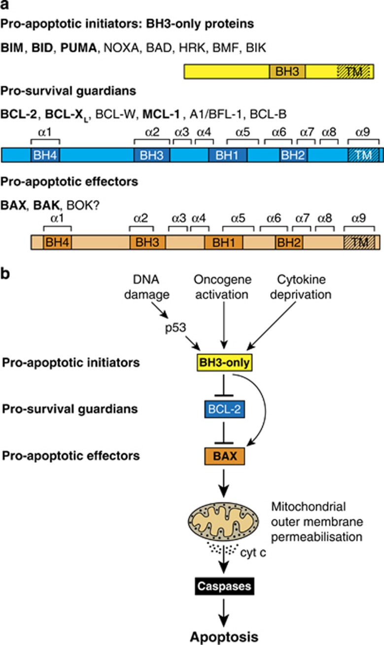
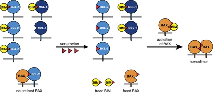
Impaired apoptosis plays a central role in cancer development and limits the efficacy of conventional cytotoxic therapies. Deepening understanding of how opposing factions of the BCL-2 protein family switch on apoptosis and of their structures has driven development of a new class of cancer drugs that targets various pro-survival members by mimicking their natural inhibitors, the BH3-only proteins. These 'BH3 mimetic' drugs seem destined to become powerful new weapons in the arsenal against cancer. Successful clinical trials of venetoclax/ABT-199, a specific inhibitor of BCL-2, have led to its approval for a refractory form of chronic lymphocytic leukaemia and to scores of on-going trials for other malignancies. Furthermore, encouraging preclinical studies of BH3 mimetics that target other BCL-2 pro-survival members, particularly MCL-1, offer promise for cancers resistant to venetoclax. This review sketches the impact of the BCL-2 family on cancer development and therapy, describes how interactions of family members trigger apoptosis and discusses the potential of BH3 mimetic drugs to advance cancer therapy.


Conflict of interest statement

WEHI has received research funding from Genentech and AbbVie, and royalty and milestone payments for venetoclax.

Figures

Figure 1
Figure 1
The BCL-2 protein family. (a) The initiator, guardian and effector factions of the family. Domains of shared BCL-2 Homology (BH), and the nine α-helices in the multi-BH domain members are indicated. (Effectors BAX and BAK, and the related BOK, have a BH4 domain if both structural and sequence homology are considered.) BOK seems to drive apoptosis only in special circumstances. Faction members most important for controlling apoptosis are in bold. All multi-BH domain family members and some BH3-only proteins (BIM, BID, BIK, HRK) have a C-terminal transmembrane (TM) domain for anchoring to organelles, most notably the MOM. (b) How the BCL-2 protein family controls cell life and death is shown. In healthy cells, the pro-survival guardians prevent activation of BAX and BAK, at least in part by binding the BH3 domain (α2) of any destabilised BAX or BAK monomers. Various stress signals activate BH3-only proteins that avidly bind their pro-survival relatives, preventing their constraint of BAX or BAK. In addition, certain BH3-only proteins, namely BIM, cleaved (active) BID and probably PUMA, can directly activate BAX and BAK, which then homo-oligomerise and permeabilise the MOM, releasing cytochrome c to initiate caspase activation and cellular demolition. Modified, with permission, from Figure 1 of Cory et al.
Figure 2
Figure 2
Interaction of BCL-2 family members. (a) The canonical BH3/surface groove interaction in the family. Structure of BCL-XL (blue surface representation) bound to the amphipathic helical BH3 peptide of BIM (a yellow ribbon indicates its helical structure) (PDB/3FDL), with its N terminus at the bottom. Underneath the protein is a consensus BH3 sequence of the pro-apoptotic proteins (x denotes nonconserved residues). The four key hydrophobic amino acids (yellow) of the Bim BH3 peptide that bind to pockets p1 to p4 in BCL-XL are highlighted, as is the invariant aspartic acid (D) (oxygens in red) that binds to a conserved arginine (R) in BCL-XL. BIM or BID BH3 peptides associate with the grooves of BAX or BAK through contacts resembling those with their pro-survival relatives (as above) but include additional contacts that contribute to their activator function., , , (b) Selective association of BH3-only proteins with their pro-survival relatives. Whereas BIM, PUMA and tBID bind promiscuously, BAD and NOXA have restricted targets, as indicated. (c) BCL-2 pro-survival targets of current BH3 mimetic drugs. A BH3 mimetic engages the surface groove of the targeted pro-survival protein(s) in a manner akin to their natural antagonists, as in (a), but usually involving only pockets p2 and p4. In cells, binding of the compound to their pro-survival target(s) releases any bound BH3-only proteins and prevents the targeted pro-survival protein(s) from restraining BAX and BAK. Note that preclinical studies were reported on Servier MCL-1 inhibitor S63845, but the clinical candidate from Novartis/Servier is the more advanced derivative S64315. Modified, with permission, from Figure 2 of Cory et al.
Figure 3
Figure 3
Model for life/death decisions on the MOM, showing how pro-survival family members constrain BAX (and BAK) and how BH3-only proteins (here BIM) drive their activation. In healthy cells, monomeric BAX shuttles between the cytosol and MOM, where VDAC2 acts as a receptor (also for BAK), although pro-survival relatives (here BCL-XL) can ‘retro-translocate’ MOM-bound BAX back to the cytosol. Upon apoptotic signalling, to allow more BAX to move to the MOM (step 1), where most BAK molecules reside, an activator BH3-only protein such as BIM may transiently engage a BAX ‘rear site’ involving helices α1 and α6,, thereby releasing the C-terminal trans-membrane (TM) domain (α9) from its surface groove to enable MOM binding. Then, groove binding by the activator drives release of the N terminus and α1 of BAX or BAK (pale orange) (step 2) and all subsequent activation steps for both the MOM-bound effector proteins. The most dramatic change is the unfolding of BAX and BAK that separates their ‘latch’ domain (α6–α8; speckled orange) from their ‘core’ domain (α2–α5), (step 3); this ejects the BH3-only activator (BIM here) and exposes the BH3 domain of BAX or BAK (α2, red triangle) (step 4). If pro-survival proteins are available to bind the exposed BAX (or BAK) BH3 domain, apoptosis aborts (step 5a). However, if pro-survival proteins are largely occupied by BH3-only proteins, the unfolded BAK or BAX monomers form homodimers through reciprocal BH3/groove interactions of their core domains (step 5b)., , , The core dimers are the central unit of the BAX and BAK homo-oligomers,, , but how they associate into oligomers (step 6) remains uncertain, as does how the oligomers drive MOM permeabilisation (see text). Modified, with permission, from Figure 3 of Cory et al.
Figure 4
Figure 4
Killing of ‘primed’ cancer cells by BH3 mimetics. Because of the many stresses imposed by the tumourigenic process, such as abridged cell cycle checkpoints, hypoxia or altered metabolism, many cancer cells have been selected for elevated expression of pro-survival proteins, here BCL-2 and MCL-1. These stresses will have elevated the level of BH3-only proteins such as BIM, much of which may be in complex with BCL-2 (top left). Hence, paradoxically, the ‘primed’ cancer cell, despite elevated BCL-2, can be nearer the apoptotic threshold than normal cell counterparts. (see text and Letai in this series.) When such cells are exposed to a BCL-2-specific BH3 mimetic like venetoclax (red triangles), BIM is displaced from BCL-2, as is any BAX previously sequestered by BCL-2 (bottom left). The freed BIM can then sequester any unoccupied non-targeted pro-survival protein (here MCL-1) (centre); it can also activate inactive BAX (or BAK) monomers (right), thereby facilitating apoptosis. Thus, the presence in a primed cancer cell of abundant complexes of a BH3-only protein like BIM with a pro-survival partner renders the cell more vulnerable to apoptosis. Once activated BAX monomers build up on the MOM, each can insert its exposed BH3 (α2) into the groove (α3–α5) of another activated monomer, generating a ‘symmetric’ homodimer (right)., , These dimers can then oligomerise and permeabilise the MOM, releasing cytochrome c (cyt c) from the intermembrane space to trigger caspase activation (see Figure 3). Modified, with permission, from Figure 5 of Cory et al.

References

    1. Croce C. miR15/16 and Bcl2: from gene discovery to treatment. Cell Death Differ 2018. 25:21–26. - PMC - PubMed
    1. Strasser A, Vaux DL. Bcl2 and cell death from an evolutionary perspective. Cell Death Differ 2018. 25:13–20. - PMC - PubMed
    1. Opferman J. BCL-2 family in development. Cell Death Differ 2018. 25:37–45. - PMC - PubMed
    1. Andrews DW. Bcl-2 family proteins: Changing partners in the dance towards death. Cell Death Differ 2018. 25:65–80. - PMC - PubMed
    1. Letai A. Why do BCL-2 inhibitors work and where should we use them in the clinic? Cell Death Differ 2018. 25:56–64. - PMC - PubMed

MeSH terms