Comparative Study
. 2018 Feb 15;197(4):481-491.
doi: 10.1164/rccm.201706-1139OC.

Mucin Production and Hydration Responses to Mucopurulent Materials in Normal versus Cystic Fibrosis Airway Epithelia

Affiliations
Comparative Study

Mucin Production and Hydration Responses to Mucopurulent Materials in Normal versus Cystic Fibrosis Airway Epithelia

Lubna H Abdullah et al. Am J Respir Crit Care Med. .

Abstract

Rationale: Cystic fibrosis (CF) airways disease produces a mucoobstructive lung phenotype characterized by airways mucus plugging, epithelial mucous cell metaplasia/hyperplasia, chronic infection, and inflammation. Simultaneous biochemical and functional in vivo studies of mucin synthesis and secretion from CF airways are not available. In vitro translational models may quantitate differential CF versus normal mucin and fluid secretory responses to infectious/inflammatory stimuli.

Objectives: We tested the hypothesis that CF airways exhibit defective epithelial fluid, but not mucin, secretory responses to bacterial/inflammatory host products.

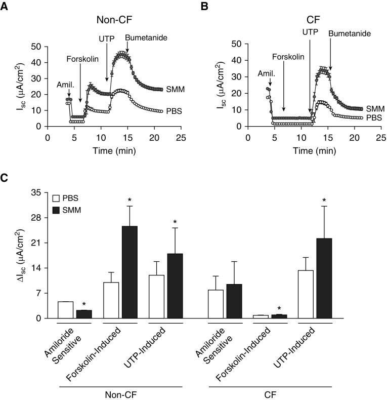
Methods: Well-differentiated primary human bronchial epithelial cultures were exposed to supernatant from mucopurulent material (SMM) from human CF airways as a test of bacterial/inflammatory host product stimulus. Human bronchial epithelia (HBE) with normal CF transmembrane conductance regulator function were compared with ΔF508/ΔF508 CF HBE.

Measurements and main results: Acute (up to 60 min) SMM exposure promoted mucin secretion, but mucins were degraded by the proteolytic enzymes present in SMM. Chronic SMM exposure induced upregulation of mucin synthesis and storage and generated absolute increases in basal and stimulated mucin release in normal and CF cultures. These responses were similar in normal and CF cultures. In contrast, SMM produced a coordinated CF transmembrane conductance regulator-mediated Cl- secretory response in normal HBE, but not in CF HBE. The absence of the fluid secretory response in CF produced quantitatively more dehydrated mucus.

Conclusions: Our study reveals the interplay between regulation of mucin and fluid secretion rates in inflamed versus noninflamed conditions and why a hyperconcentrated mucus is produced in CF airways.

Keywords: airway hydration; airway inflammation; airway mucins; cystic fibrosis.


Figures

Figure 1.
Figure 1.
Acute supernatant of mucopurulent material (SMM) exposure induces mucin release and promotes degradation of secreted mucins. After washing away accumulated mucins, non–cystic fibrosis (normal) human bronchial epithelial cultures were mucosally exposed to various doses of SMM for 1 hour. Secreted mucins were collected from the epithelial surface and the cultures were fixed. (A) Alcian blue/periodic acid–Schiff (AB/PAS) staining of human bronchial epithelial cultures exposed to phosphate-buffered saline (control), 100-μM ATPγS, or various doses of SMM. ATPγS was used as a positive control for mucin release. (B) Quantification of the dose-dependent effect of acute SMM-promoted mucin exocytosis. (C) Secreted mucin levels (measured by ELISA) from the cultures depicted in A. Data represent mean ± SD; n = 4–5. *P < 0.05, ATPγS-treated or SMM-exposed versus control cultures. Scale bar = 20 μm. ATPγS = adenosine 5′-O-(3-thio)triphosphate.
Figure 2.
Figure 2.
Time course for acute supernatant of mucopurulent material (SMM)-induced mucin release and degradation of secreted mucins. (A) Alcian blue/periodic acid–Schiff (AB/PAS) staining of non–cystic fibrosis (normal) human bronchial epithelial cultures mucosally exposed to undiluted SMM for various amounts of time up to 60 minutes. (B) Quantification of the time-dependent effect of acute SMM-promoted mucin exocytosis. (C) Secreted mucin levels (measured by ELISA) from the cultures depicted in A. Apical secretions were collected in buffer containing protease and elastase inhibitors. Cultures were fixed immediately after sample collection. Depletion of mucin stores occurs as early as 5 minutes and is almost complete by 60 minutes. Approximately 60% of secreted mucins are degraded in 1 hour, as compared with secreted mucins after 5-minute SMM exposure. Data represent mean ± SD. n = 5. *P < 0.05, SMM-exposed versus control cultures. Scale bar = 20 μm.
Figure 3.
Figure 3.
Protease inhibition prevents mucin degradation by supernatant of mucopurulent material (SMM). Mucins harvested from non–cystic fibrosis (normal) human bronchial epithelia were incubated with different doses of SMM in presence or absence of protease inhibitors (PI) for 30 minutes. Samples were evaluated for MUC5B, because it is the major mucin produced and secreted by human bronchial epithelial cultures. Data represent mean ± SD. n = 3. *P < 0.05, SMM-exposed versus control cultures not incubated with PI.
Figure 4.
Figure 4.
Dose–response for chronic supernatant of mucopurulent material (SMM) exposure on mucin gene expression. Non–cystic fibrosis (normal) human bronchial epithelial cultures were washed and mucosally exposed to various doses of SMM for 48 hours. Cultures were washed again and extracted in RNeasy guanidine-based buffer, and MUC5AC and MUC5B mRNA levels were determined by qRT-PCR. Data represent mean ± SD. n = 6. *P < 0.05, SMM-exposed versus control cultures.
Figure 5.
Figure 5.
Dose–response for chronic supernatant of mucopurulent material (SMM) exposure on MUC5AC and MUC5B protein synthesis. Non–cystic fibrosis (normal) human bronchial epithelial cultures were washed and mucosally exposed to different dilutions of SMM for 48 hours. Cultures were extracted in RNeasy guanidine-based buffer, and aliquots electrophoresed in agarose. Immunoblots were probed with antibodies to the immature and mature forms of MUC5AC (A and B) and MUC5B (C and D). Data represent mean ± SD. n = 5–6. *P < 0.05, SMM-exposed versus control cultures.
Figure 6.
Figure 6.
Effect of chronic (72 h) supernatant of mucopurulent material (SMM) treatment on MUC5AC and MUC5B mRNA and protein levels. Non–cystic fibrosis (normal) human bronchial epithelial cultures were washed and mucosally exposed to undiluted SMM for 72 hours. Cultures were extracted in RNeasy guanidine-based buffer. Aliquots were either used for determination of MUC5AC and MUC5B mRNA levels by qRT-PCR (A) or electrophoresed in agarose for evaluation of the immature and mature forms of MUC5AC (B) and MUC5B (C). (D) Compiled data expressed as fold change of SMM/phosphate-buffered saline from the experiments illustrated in B and C. Data represent mean ± SD. n = 6–8. *P < 0.05, SMM-exposed versus control (phosphate-buffered saline) cultures.
Figure 7.
Figure 7.
Chronic (72 h) supernatant of mucopurulent material (SMM) treatment of non–cystic fibrosis (CF) and CF human bronchial epithelial cultures promotes increased mucin secretion. Accumulated secreted mucins and ATPγS (100 μM)-induced release of intracellular stored mucins from non-CF (normal) and CF human bronchial epithelial cultures exposed to phosphate-buffered saline (PBS) (control) or undiluted SMM for 72 hours. The apically accumulated mucins were assayed by ELISA, using mucin-specific antibodies, as detailed in Methods. Data represent mean ± SD. n = 5. *P < 0.05, SMM-exposed non-CF or CF versus control (PBS-treated) cultures. #P < 0.05, CF versus non-CF cultures exposed to PBS. ATPγS = adenosine 5′-O-(3-thio)triphosphate.
Figure 8.
Figure 8.
Effect of supernatant of mucopurulent material (SMM) on the bioelectric properties of non–cystic fibrosis (CF) and CF human bronchial epithelia. Bioelectric measurements were conducted in non-CF (normal) and CF human bronchial epithelial cultures, as described in Methods. (A and B) Effect of sequential addition of amiloride (Amil.) (300 μM; apical), forskolin (10 μM; apical), UTP (100 μM; apical), and bumetanide (100 μM; basolateral) on short circuit currents in non-CF (A) and CF (B) cultures exposed to phosphate-buffered saline (PBS) or SMM for 72 hours. (C) Compiled data from A and B for amiloride-sensitive and forskolin- and UTP-induced currents. Data represent mean ± SD. n = 8–12. *P < 0.05, SMM-exposed versus control (PBS-exposed) cultures. ISC = short circuit current; UTP = uridine-5′-triphosphate.
Figure 9.
Figure 9.
Effect of supernatant of mucopurulent material (SMM) on airway surface liquid (ASL) height in non–cystic fibrosis (CF) versus CF human bronchial epithelial cultures. (A) Non-CF (normal) and CF human bronchial epithelial cultures were exposed to phosphate-buffered saline (PBS) or SMM for 72 hours. ASL height was evaluated as described in Methods, and is shown as the percentage increase after treatment with SMM over PBS for both groups. Data represent mean ± SD. n = 5–6. *P < 0.05, CF versus non-CF cultures. (B) Non-CF human bronchial epithelial cultures were exposed to SMM for 48 hours and ASL height was evaluated by XZ confocal microscopy. CFTRInh-172 was added apically as a dry powder in perfluorocarbon and ASL height was remeasured. ASL height is expressed as percentage inhibition of ASL height by CFTRInh-172 relative to ASL height from PBS- or SMM-treated non-CF human bronchial epithelia. Data represent mean ± SD. n = 4. *P < 0.05, SMM + CFTRInh-172 versus SMM. CFTR = CF transmembrane conductance regulator.
Figure 10.
Figure 10.
Effect of supernatant of mucopurulent material (SMM) on mucus percent solids in non–cystic fibrosis (CF) versus CF human bronchial epithelial cultures. Non-CF (normal) and CF cultures were exposed to phosphate-buffered saline or SMM for 72 hours. Mucus percent solids was evaluated as described in Methods. Data represent mean ± SD. n = 8 for all groups. *P < 0.05, CF versus non-CF cultures. PBS = phosphate-buffered saline.

Comment in

References

    1. Knowles MR, Boucher RC. Mucus clearance as a primary innate defense mechanism for mammalian airways. J Clin Invest. 2002;109:571–577. - PMC - PubMed
    1. Kesimer M, Ehre C, Burns KA, Davis CW, Sheehan JK, Pickles RJ. Molecular organization of the mucins and glycocalyx underlying mucus transport over mucosal surfaces of the airways. Mucosal Immunol. 2013;6:379–392. - PMC - PubMed
    1. Holmén JM, Karlsson NG, Abdullah LH, Randell SH, Sheehan JK, Hansson GC, et al. Mucins and their O-glycans from human bronchial epithelial cell cultures. Am J Physiol Lung Cell Mol Physiol. 2004;287:L824–L834. - PubMed
    1. Abdullah LH, Davis SW, Burch L, Yamauchi M, Randell SH, Nettesheim P, et al. P2u purinoceptor regulation of mucin secretion in SPOC1 cells, a goblet cell line from the airways. Biochem J. 1996;316:943–951. - PMC - PubMed
    1. Rose MC, Voynow JA. Respiratory tract mucin genes and mucin glycoproteins in health and disease. Physiol Rev. 2006;86:245–278. - PubMed

Publication types