Review
. 2017 Nov 3;18(11):2315.
doi: 10.3390/ijms18112315.

Oxidative Stress in Human Atherothrombosis: Sources, Markers and Therapeutic Targets

Affiliations
Review

Oxidative Stress in Human Atherothrombosis: Sources, Markers and Therapeutic Targets

Jose Luis Martin-Ventura et al. Int J Mol Sci. .

Abstract

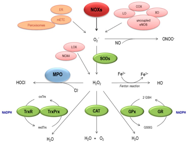
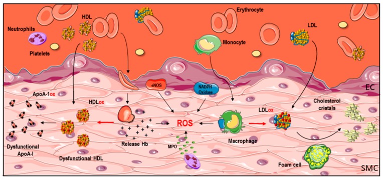
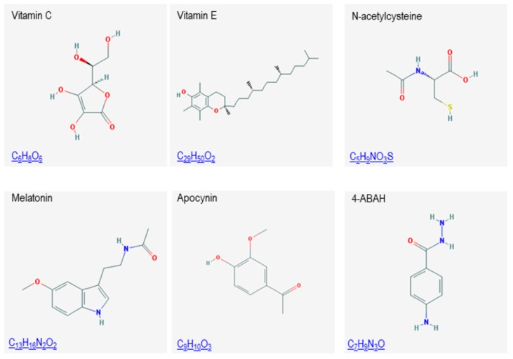
Atherothrombosis remains one of the main causes of morbidity and mortality worldwide. The underlying pathology is a chronic pathological vascular remodeling of the arterial wall involving several pathways, including oxidative stress. Cellular and animal studies have provided compelling evidence of the direct role of oxidative stress in atherothrombosis, but such a relationship is not clearly established in humans and, to date, clinical trials on the possible beneficial effects of antioxidant therapy have provided equivocal results. Nicotinamide adenine dinucleotide phosphate (NADPH) oxidase is one of the main sources of reactive oxygen species (ROS) in human atherothrombosis. Moreover, leukocyte-derived myeloperoxidase (MPO) and red blood cell-derived iron could be involved in the oxidative modification of lipids/lipoproteins (LDL/HDL) in the arterial wall. Interestingly, oxidized lipoproteins, and antioxidants, have been analyzed as potential markers of oxidative stress in the plasma of patients with atherothrombosis. In this review, we will revise sources of ROS, focusing on NADPH oxidase, but also on MPO and iron. We will also discuss the impact of these oxidative systems on LDL and HDL, as well as the value of these modified lipoproteins as circulating markers of oxidative stress in atherothrombosis. We will finish by reviewing some antioxidant systems and compounds as therapeutic strategies to prevent pathological vascular remodeling.

Keywords: atherothrombosis; biomarkers; lipids/lipoprotein oxidation; oxidative stress.


Conflict of interest statement

The authors declare no conflict of interest.

Figures

Figure 1
Figure 1
Generation and elimination of ROS. Enzymatic systems (in red) including NADPH oxidases (NOXs), Xanthine Oxidase (XO), Lipoxygenase (LO), Cyclooxygenase (COX) and uncoupled eNOS produce O2 that can also be generated non-enzymatically (in orange) by the mitochondrial electron transport chain (mETC), the endoplasmic reticulum (ER) and peroxisomes. O2 is then transformed into H2O2 spontaneously or through superoxide dismutases (SODs) or can be synthesized directly by NOX-4 or as a by-product of lysyl oxidase (LOX). O2 can rapidly react with NO leading to the formation of ONOO. H2O2 can be then converted into more reactive molecules, including hydroxyl radical (OH) by Fenton reaction or into HOCl by myeloperoxidase (MPO). Furthermore, H2O2 can also be transformed into H2O by catalase (CAT) or by the glutathione peroxidase (GPx)/gluthathione reductase (GR) and the thioredoxin (Trx)/peroxiredoxin (PRx) systems. TrxR: thioredoxin reductase; TrxPrx: thioredoxin peroxidase.
Figure 2
Figure 2
Sources of oxidative stress in the vascular wall. The oxidative process inside the pathological vascular wall is the result of the interaction of lipids/lipoproteins and reactive oxygen species (ROS) derived from infiltrating (red blood cells-RBC, platelets, leukocytes-neutrophils and monocytes) and resident (endothelial cells-EC- and smooth muscle cells-SMC-) cells. LDL, low-density lipoproteins; HDL, high-density lipoproteins; ApoA1, apolipoprotein A1; MPO, myeloperoxidase; Hb, hemoglobin; eNOS, endothelial NO synthase. Some graphical elements from this figure were adapted from Servier Medical Art Powerpoint image bank at http://smart.servier.com/.
Figure 3
Figure 3
Chemical structures of different antioxidant compounds.

Similar articles

Cited by

References

    1. Bentzon J.F., Otsuka F., Virmani R., Falk E. Mechanisms of plaque formation and rupture. Circ. Res. 2014;114:1852–1866. doi: 10.1161/CIRCRESAHA.114.302721. - DOI - PubMed
    1. Weintraub N.L. Understanding abdominal aortic aneurysm. N. Engl. J. Med. 2009;361:1114–1116. doi: 10.1056/NEJMcibr0905244. - DOI - PMC - PubMed
    1. Chen Q., Wang Q., Zhu J., Xiao Q., Zhang L. Reactive oxygen species: Key regulators in vascular health and diseases. Br. J. Pharmacol. 2017 doi: 10.1111/bph.13828. - DOI - PMC - PubMed
    1. Montezano A.C., Dulak-Lis M., Tsiropoulou S., Harvey A., Briones A.M., Touyz R.M. Oxidative stress and human hypertension: Vascular mechanisms, biomarkers, and novel therapies. Can. J. Cardiol. 2015;31:631–641. doi: 10.1016/j.cjca.2015.02.008. - DOI - PubMed
    1. Konior A., Schramm A., Czesnikiewicz-Guzik M., Guzik T.J. NADPH oxidases in vascular pathology. Antioxid. Redox Signal. 2014;20:2794–2814. doi: 10.1089/ars.2013.5607. - DOI - PMC - PubMed

MeSH terms