. 2017 Nov 3;12(11):e0186899.
doi: 10.1371/journal.pone.0186899. eCollection 2017.

Water without windows: Evaluating the performance of open cell transmission electron microscopy under saturated water vapor conditions, and assessing its potential for microscopy of hydrated biological specimens

Affiliations

Water without windows: Evaluating the performance of open cell transmission electron microscopy under saturated water vapor conditions, and assessing its potential for microscopy of hydrated biological specimens

Cathal Cassidy et al. PLoS One. .

Abstract

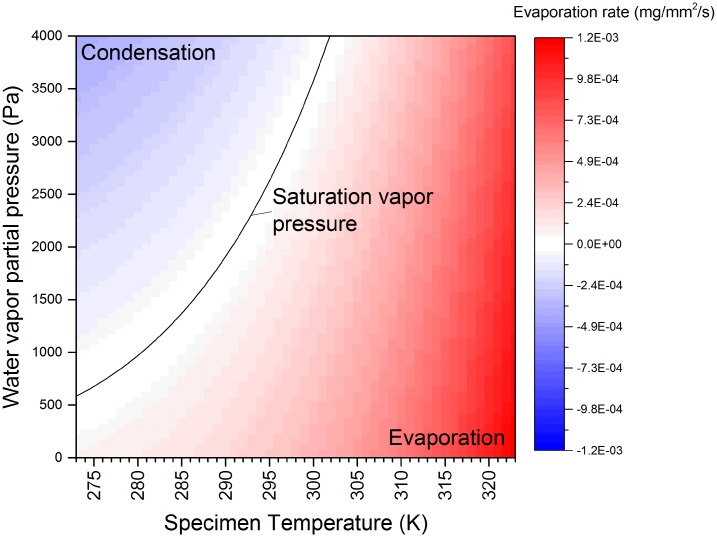
We have performed open cell transmission electron microscopy experiments through pure water vapor in the saturation pressure regime (>0.6 kPa), in a modern microscope capable of sub-Å resolution. We have systematically studied achievable pressure levels, stability and gas purity, effective thickness of the water vapor column and associated electron scattering processes, and the effect of gas pressure on electron optical resolution and image contrast. For example, for 1.3 kPa pure water vapor and 300kV electrons, we report pressure stability of ± 20 Pa over tens of minutes, effective thickness of 0.57 inelastic mean free paths, lattice resolution of 0.14 nm on a reference Au specimen, and no significant degradation in contrast or stability of a biological specimen (M13 virus, with 6 nm body diameter). We have also done some brief experiments to confirm feasibility of loading specimens into an in situ water vapor ambient without exposure to intermediate desiccating conditions. Finally, we have also checked if water experiments had any discernible impact on the microscope performance, and report pertinent vacuum and electron optical data, for reference purposes.


Conflict of interest statement

Competing Interests: The authors have declared that no competing interests exist.

Figures

Fig 1
Fig 1. Net evaporation as a function of temperature and pressure, plotted using Eq 3.
Evaporation rate per unit time and surface area, as a function of ambient water vapor partial pressure and specimen temperture (gas ambient temperature set to 298 K). The microscope used for this work allowed an ambient pressure of up to 2 kPa, indicating that the zero net evaporation condition is within range (with appropriate specimen cooling).
Fig 2
Fig 2. Schematic diagram (top) and photographs (bottom) of the experimental set-up.
The acronyms in the schematic diagram refer to: TMP (Turbo Molecular Pump), RP (Roughing pump), RGA (Residual Gas Analyser), BC (Barocel pressure gauge), DV (Diaphragm valve), NV (Needle valve), OL (Objective lens).
Fig 3
Fig 3. Gas composition and pressure control.
A. RGA spectrum acquired at a specimen area pressure of 1.98 kPa (room temperature). Most of the detected ion current is associated with water vapor (at molecular mass 18) and its ionization fragments. Small peaks from residual nitrogen, oxygen and carbon dioxide are also detected. B. Curves demonstrating the system control over the gas pressure. We show the optimum stability of the system when the set-up parameters are fixed (no fluctuations are detected, within the measurement sensitivity of the gauge, over periods exceeding twenty minutes). We also show the response under fine adjustment of the inlet needle valve, demonstrating fine control and identifying that the smallest measurable pressure shift is ∼20 Pa. We also demonstrate coarse adjustment, to a defined setpoint over a wide pressure differential, demonstrating the coarse control, transition time and stability after adjustment.
Fig 4
Fig 4. Zero-loss and low-loss EELS spectra of water vapor at various pressures (300kV, room temperature).
The zero-loss-peak FWHM is approximately 1 eV, and the observed loss peaks are centered at 7.7 eV, 10.1 eV, 13.6 eV and 18.1 eV. These values are consistent with prior reports on ultraviolet absorption edges in water vapor (see main text). Zero-loss peak intensity decreases, and energy loss intensity increases, as a function of water vapor pressure, as expected. The integration times were 1ms (zero-loss region) and 200ms (low-loss region), respectively.
Fig 5
Fig 5. Time-resolved EELS spectra, as a function of decreasing water vapor pressure.
Zero-loss and low-loss EELS spectra were acquired as a function of time, during a gas pressure skew, reducing to vacuum levels from an initial pressure of 1.4 kPa. The exact pressure skew is included in S4 Fig. The column valve open/close events allow the EELS spectra and pressure gauge readings to be precisely synchronized.
Fig 6
Fig 6. Effective thickness of water vapor for 300 kV electrons, expressed in terms of t/λ, as a function of indicated pressure.
Effective thickness has been calculated according to (tλ)P=lnIVacdEIZLP(P)dE, with full details in S3 Appendix. The effective thickness is quite linear as a function of pressure. For comparison purposes, we have also included the equivalent thickness of amorphous ice (assuming an inelastic mean free path of 330 nm at 300 kV [28]. We indicate (*) a literature value for the effective thickness of enclosing SiN membranes in a conventional closed liquid cell, from [27]. We have also marked (#) the effective water vapor thickness corresponding to saturation vapor pressure at 277 K (0.32 at 729 Pa).
Fig 7
Fig 7. Microscope resolution as a function of water vapor pressure in the specimen area.
Scale bars are 10 nm, 2 nm, 10 nm−1 and 10 nm−1, respectively. The specimen is polycrystalline gold on a carbon support. We observed maximum lattice resolution of 0.1 nm from Au(400), 0.12 nm from Au(222), and 0.14 nm from Au(220), and Young’s fringes resolution of 0.11 nm, 0.14 nm and 0.2 nm, at water vapor pressures of <1 mPa (vacuum), 0.65 kPa and 1.3 kPa, respectively. Atomic resolution is readily attained over this water vapor pressure range.
Fig 8
Fig 8. Biological specimen contrast when imaged through water vapor in the saturation pressure regime.
A. M13 phage (unstained), imaged at 300 kV in high vacuum and in 1.3 kPa water vapor ambient, at magnifications of 10kx, 43kx and 115kx (Scale bars are 500 nm, 200 nm, 50 nm, respectively). B. Sketch of M13 phage structure, based upon that described in [31]. C. Line profiles extracted across several individual filamentous phages, for the vacuum and 1.3 kPa ambient cases. Individual profile locations and data are included in S2 Dataset.
Fig 9
Fig 9. Demonstration of feasibility of specimen loading into a water vapor ambient in the microscope, without exposure to desiccating conditions.
We confirmed a loading sequence that allowed the specimen to be loaded into the gas-laden microscope, without exposure to intermediate high vacuum conditions. Letters A-L and P-T are referred to in the text. It took approximately 5 minutes to get the specimen, sealed in the internal holder cavity, from the bench into preset gas-laden microscope.
Fig 10
Fig 10. Upper column and electron gun vacuum levels during specimen area water vapor exposures (column valves open).
As expected, the correlation between with specimen area pressure and the gun area pressure weakens closer to the electron gun. There was no detectable shift in inner gun vacuum readings, with maximum (kPa) levels of water vapor in the specimen area. The labels BC/O, IGPc1, IGPa and IGPf refer to the specific microscope hardware used.
Fig 11
Fig 11. Post-water vapor vacuum logging and aberration measurement.
A. After execution of water vapor experiments, the system was typically left pumping overnight, and had fully recovered to normal vacuum levels by the following morning. B. Electron optical aberrations, as measured using the Cs-corrector hardware, showed no shift as a function of water vapor exposure.

References

    1. Jinschek JR. Advances in the environmental transmission electron microscope (ETEM) for nanoscale in situ studies of gas-solid interactions. Chemical Communications. 2014;50:2696–2706. doi: 10.1039/C3CC49092K - DOI - PubMed
    1. Ek M, Jespersen SPF, Damsgaard CD, Helveg S. On the role of the gas environment, electron-dose-rate, and sample on the image resolution in the transmission electron microscope. Advanced Structural and Chemical Imaging. 2016;2(4):1–8.
    1. Cameron RE, McDonald AM. Minimizing sample evaporation in the environmental scanning electron microscope. Journal of Microscopy. 1994;173(3):227–237. doi: 10.1111/j.1365-2818.1994.tb03445.x - DOI
    1. Ross FM, Wang C, de Jonge N. Transmission electron microscopy of specimens and processes in liquids. MRS Bulletin. 2016;41:791–799. doi: 10.1557/mrs.2016.212 - DOI
    1. Ross FM. Opportunities and challenges in liquid cell electron microscopy. Science. 2015;350(6267):aaa9886–1–aaa9886–9. doi: 10.1126/science.aaa9886 - DOI - PubMed

MeSH terms

LinkOut - more resources