. 2017 Dec;128(12):2470-2481.
doi: 10.1016/j.clinph.2017.08.026. Epub 2017 Sep 19.

Toward noninvasive monitoring of ongoing electrical activity of human uterus and fetal heart and brain

Affiliations

Toward noninvasive monitoring of ongoing electrical activity of human uterus and fetal heart and brain

S Lew et al. Clin Neurophysiol. 2017 Dec.

Abstract

Objective: To evaluate whether a full-coverage fetal-maternal scanner can noninvasively monitor ongoing electrophysiological activity of maternal and fetal organs.

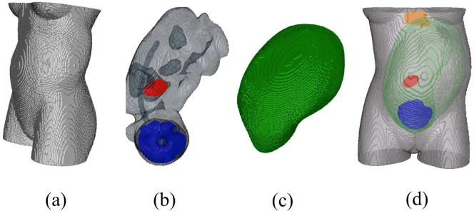
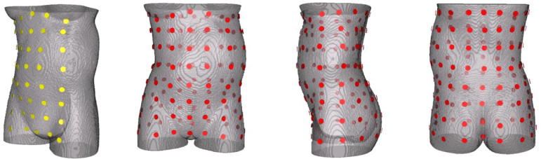
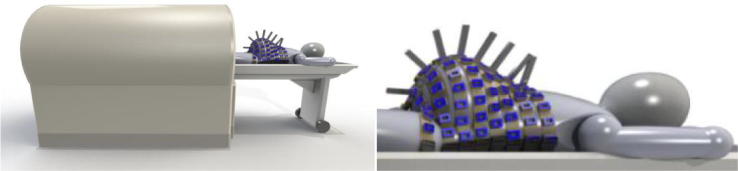
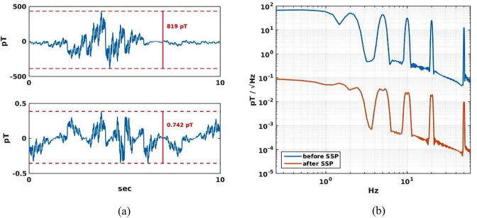
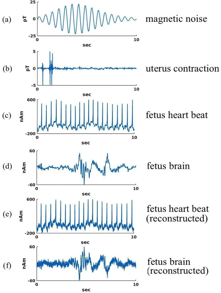
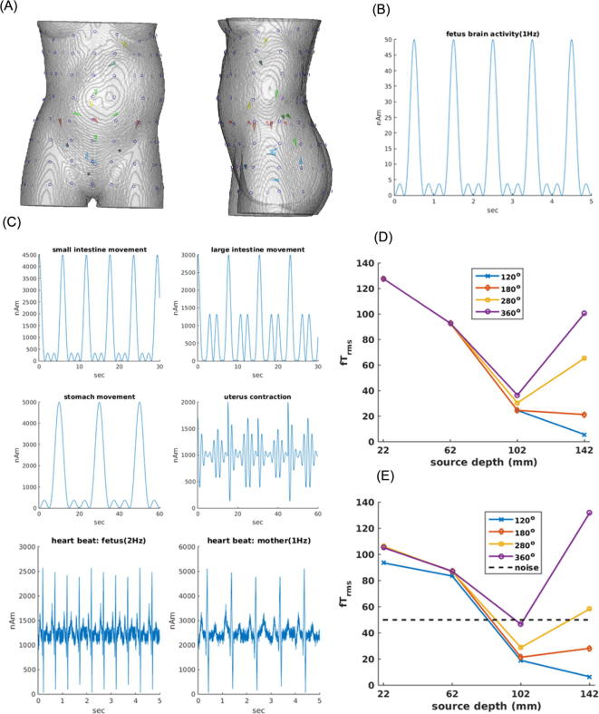
Methods: A simulation study was carried out for a scanner with an array of magnetic field sensors placed all around the torso from the chest to the hip within a horizontal magnetic shielding enclosure. The magnetic fields from internal organs and an external noise source were computed for a pregnant woman with a 35-week old fetus. Signal processing methods were used to reject the external and internal interferences, to visualize uterine activity, and to detect activity of fetal heart and brain.

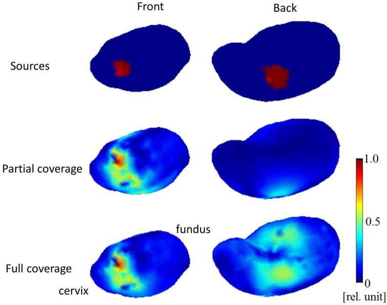
Results: External interference was reduced by a factor of 1000, sufficient for detecting signals from internal organs when combined with passive and active shielding. The scanner rejects internal interferences better than partial-coverage arrays. It can be used to estimate currents around the uterus. It clearly detects spontaneous activity from the fetal heart and brain without averaging and weaker evoked brain activity at all fetal head positions after averaging.

Conclusion: The simulated device will be able to monitor the ongoing activity of the fetal and maternal organs.

Significance: This type of scanner may become a novel tool in fetal medicine.

Keywords: Electrocardiography (ECG); Electroencephalography (EEG); Electrohysterography (EHG); Magnetocardiography (MCG); Magnetoencephalography (MEG); Prenatal medicine.


Figures

Fig. 1
Fig. 1
The compartments of the FEM model: (a) torso (b) fetus brain (blue), eyes, heart (red), lungs, stomach, bladder, (c) uterus, (d) torso with fetus brain, fetus heart, uterus, and mother heart (orange). (For interpretation of the references to colour in this figure legend, the reader is referred to the web version of this article.)
Fig. 2
Fig. 2
(Left) Partial-coverage sensor array (8 × 5 array, 40 channels). (Right) Full-coverage sensor array (8 × 16 array, 128 channels).
Fig. 3
Fig. 3
Initial design of the FM scanner with a full-coverage sensor array consisting of OPM sensors inserted into the light-weight, flexible belts placed around the body of the mother. Courtesy of Anthony Mascarenas, Tristan Technologies, Inc., San Diego, CA.
Fig. 4
Fig. 4
Rejection of external magnetic disturbance. (a) External noise field before (top) and after (bottom) SSP. (b) The same noise field presented in frequency domain.
Fig. 5
Fig. 5
Subspace angle (ϖ) between the signal dipole (red sphere) and noise dipole 12 as a function of coverage angle of the sensor array (Ω) for different angular separations θ (angle between + y axis and the dipole projected on the yz plane) and ϕ (angle between + x and the dipole projected on xz plane). For this shallow noise dipole below the sensor array with the smallest coverage angle, ϖ is independent of Ω, but depends on θ and ϕ. Top inset shows the location of the signal dipole in the fetal brain (red sphere), pointing to feet, and 23 noise dipoles inside the torso.
Fig. 6
Fig. 6
Subspace angle (ϖ) between the signal dipole (red sphere (see Fig. 5 inset)) and noise dipole 5 as a function of coverage angle of the sensor array (Ω) for different angular separations θ and ϕ. For this deep and lateral noise dipole, ϖ depends on Ω, θ and ϕ.
Fig. 7
Fig. 7
Imaging of the myogenic current in the entire uterus with the partial and full coverage sensor arrays.
Fig. 8
Fig. 8
Extraction of fetal heart and brain activity in the presence of an external noise source and internal interference sources. (a) External disturbance. (b) Uterine contraction signal. (c) Fetal heart signal. (d) Fetal brain signal. (e) Extracted fetal heart activity. (f) Extracted fetal brain activity.
Fig. 9
Fig. 9
Magnetic field rms strength for a current dipole in the fetal brain as a function of the dipole depth and sensor coverage angle with and without interfering magnetic field noise from internal organs. (A) Signal dipole – a single dipole in the fetal brain (red line with a circle) located at 4 positions along the midline close to the axis passing through the navel. Noise dipoles - a single noise dipole located in the maternal and another in the fetal heart (yellow arrows), a single dipole in the maternal stomach (green, just below the maternal heart source), 5 dipoles in the uterus near the cervix (blue arrows), 2 dipoles in the large intestines (brown arrows on the left and right side on the plane of the fetal brain dipole), and 4 dipoles in the small intestines (light green arrows on a posterior plane centered on the fetal brain dipole). (B) Temporal waveform of the fetal brain dipole (50 nAm maximum). (C) Temporal waveforms of the noise dipoles in the internal organs. Note their moments are 2000–6000 nAm, about 100× stronger than the moment of the fetal brain dipole. (D) Maximum rms field strength at the sensor array for the fetal brain dipole located at 4 depths without any noise field from the internal organs. (E) Same with the interfering magnetic field from the internal organs. Dotted line indicates the noise level of the simulated OPM sensors in fTrm. (For interpretation of the references to colour in this figure legend, the reader is referred to the web version of this article.)

Comment in

Similar articles

Cited by

References

    1. Alamedine D., Khalil M., Marque C. Comparison of different EHG feature selection methods for the detection of preterm labor. Comput Math Methods Med. 2013;2013:485684. - PMC - PubMed
    1. Alem O., Sander T.H., Mhaskar R., LeBlanc J., Eswaran H., Steinhoff U., Okada Y., Kitching J., Trahms L., Knappe S. Fetal magnetocardiography measurements with an array of microfabricated optically pumped magnetometers. Phys Med Biol. 2015;60:4797–4811. - PubMed
    1. Alexandersson A., Steingrimsdottir T., Terrien J., Marque C., Karlsson B. The Icelandic 16-channel electrode electrohysterogram database. Sci Data. 2015;2:150017. - PMC - PubMed
    1. Bakker J.J.H., Verhoeven C.J.M., Janssen P.F., van Lith J.M., van Uodgaarden E.D., Boemenkamp K.W.M., Pasatsonis D.N.M., Willem B., Mol J., van der Post J.A.M. Outcomes after internal versus external tocodynamometry for monitoring labor. New Engl J Med. 2010;362:306–313. - PubMed
    1. Bibin L., Anquez J., Alcalde J.P., Boubekeur T., Angelini E., Bloch I. Whole body pregnant woman modeling by digital geometry processing with detailed utero-fetal unit based on medical images. IEEE Trans Biomed Eng. 2010;57:2346–2358. - PubMed

Publication types

LinkOut - more resources