. 2017 Nov 2;101(5):716-724.
doi: 10.1016/j.ajhg.2017.09.014.

De Novo Missense Mutations in DHX30 Impair Global Translation and Cause a Neurodevelopmental Disorder

Affiliations

De Novo Missense Mutations in DHX30 Impair Global Translation and Cause a Neurodevelopmental Disorder

Davor Lessel et al. Am J Hum Genet. .

Erratum in

  • De Novo Missense Mutations in DHX30 Impair Global Translation and Cause a Neurodevelopmental Disorder.
    Lessel D, Schob C, Küry S, Reijnders MRF, Harel T, Eldomery MK, Coban-Akdemir Z, Denecke J, Edvardson S, Colin E, Stegmann APA, Gerkes EH, Tessarech M, Bonneau D, Barth M, Besnard T, Cogné B, Revah-Politi A, Strom TM, Rosenfeld JA, Yang Y, Posey JE, Immken L, Oundjian N, Helbig KL, Meeks N, Zegar K, Morton J, The Ddd Study, Schieving JH, Claasen A, Huentelman M, Narayanan V, Ramsey K; C4RCD Research Group; Brunner HG, Elpeleg O, Mercier S, Bézieau S, Kubisch C, Kleefstra T, Kindler S, Lupski JR, Kreienkamp HJ. Lessel D, et al. Am J Hum Genet. 2018 Jan 4;102(1):196. doi: 10.1016/j.ajhg.2017.12.016. Am J Hum Genet. 2018. PMID: 29304375 Free PMC article. No abstract available.

Abstract

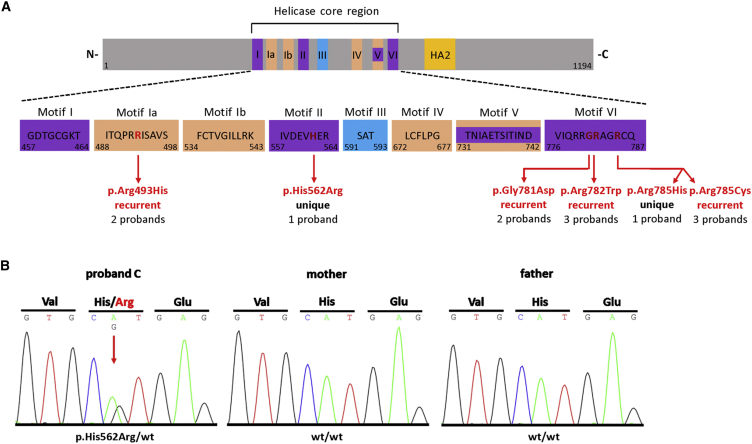
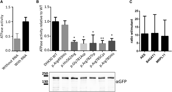
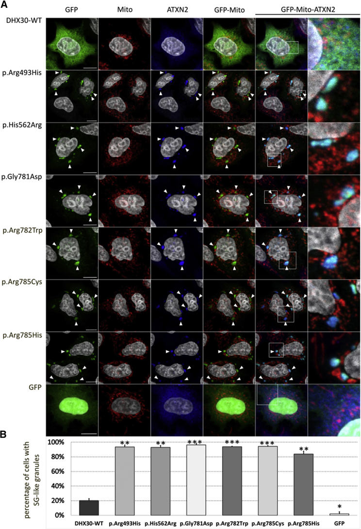
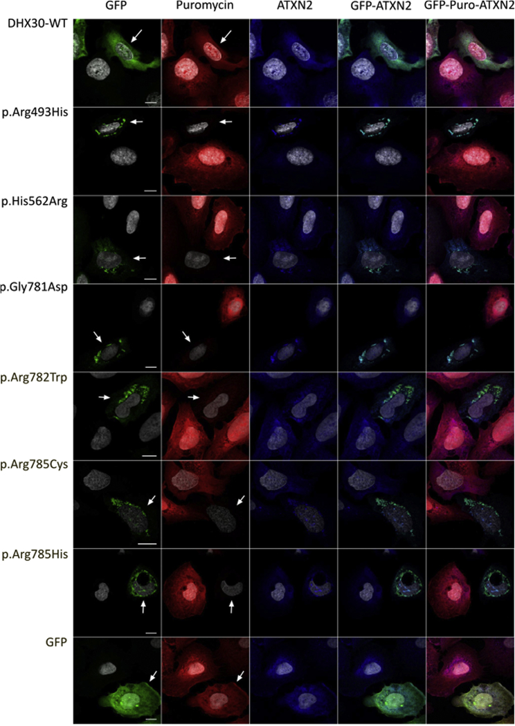
DHX30 is a member of the family of DExH-box helicases, which use ATP hydrolysis to unwind RNA secondary structures. Here we identified six different de novo missense mutations in DHX30 in twelve unrelated individuals affected by global developmental delay (GDD), intellectual disability (ID), severe speech impairment and gait abnormalities. While four mutations are recurrent, two are unique with one affecting the codon of one recurrent mutation. All amino acid changes are located within highly conserved helicase motifs and were found to either impair ATPase activity or RNA recognition in different in vitro assays. Moreover, protein variants exhibit an increased propensity to trigger stress granule (SG) formation resulting in global translation inhibition. Thus, our findings highlight the prominent role of translation control in development and function of the central nervous system and also provide molecular insight into how DHX30 dysfunction might cause a neurodevelopmental disorder.


Figures

Figure 1
Figure 1
Identified Variants Are Localized within Conserved Helicase Motifs of DHX30 (A) Top: Schematic protein structure of DHX30 showing conserved motifs of the helicase core region and the helicase associated domain (HA2). Nucleotide-interacting motifs (I, II, and VI) are shown in purple, nucleic acid-binding motifs (Ia, Ib, and IV) in orange, motif V, which binds nucleic acid and interacts with nucleotides, in purple and orange, and motif III, which couples ATP hydrolysis to RNA unwinding, in blue. (N- N terminus; C- C terminus). Bottom: Amino acids within conserved motifs of the helicase core region. The position of the first and last amino acid within each motif is denoted below left and right, respectively. The position of the de novo mutations identified in this study are marked with vertical red arrows and shown in red. (B) Sanger sequence electropherograms of parts of DHX30 after PCR amplification of genomic DNA of the affected individual C and his parents, exemplifying de novo status of the mutations identified here. The amino acid translation is shown in the three-letter code above the DNA sequence. The red arrow indicates the heterozygous mutation at c.1685A>G, (p.His562Arg), present only in the DNA sample of the affected individual.
Figure 2
Figure 2
Recombinant Protein Variants of DHX30 Affect Either ATPase Activity or RNA-Binding (A) GFP-tagged DHX30-WT was immunoprecipitated from HEK293T cell lysates using GFP-Trap_A matrix and assayed for ATPase activity first in the absence and then in the presence of exogenous RNA. Values are normalized on ATPase activity obtained with RNA. (B) ATPase assays were repeated for WT and protein variants of GFP-DHX30 in the presence of RNA. In each case, ATPase activity was normalized to the amount of DHX30 protein, as determined by western blotting using anti-GFP. ,∗∗: significantly different from DHX30-WT (p < 0.05; ∗∗p < 0.01; n = 4; ANOVA, followed by Dunnett’s multiple comparisons test). (C) RNAs extracted from GFP-Trap_A precipitates of DHX30-WT, p.Arg493His, and GFP-mCherry fusion proteins obtained from transfected HEK293T cells were subjected to gene-expression analysis using TaqMan probes for specific human mRNAs. The bar graph displays the fold enrichment of AES, B4GAT1, and MRPL11 transcripts, respectively, in DHX30-WT compared to p.Arg493His precipitates. Vertical lines indicate SD.
Figure 3
Figure 3
Recombinant Protein Variants of DHX30 Initiate the Formation of SG-like Cytoplasmic Aggregates (A) Immunocytochemical detection of DHX30-GFP fusion proteins (GFP, green), mitochondria (Mito, red), and endogenous ATXN2 (blue) in transfected U2OS cells. Regions shown at high magnification in the rightmost panels are indicated by boxes. Whereas wild-type DHX30-GFP preferentially resides throughout the cytoplasm and GFP accumulates in nuclei, recombinant protein variants of DHX30 induce the genesis of cytoplasmic foci containing endogenous SG-marker ATXN2 (arrowheads). Nuclei are identified via DAPI staining (gray). (B) Bar graph indicating the percentage of transfected cells, in which recombinant GFP proteins induce the emergence of SG-like structures. Vertical bars indicate SD. Statistical analysis was performed using unpaired Student’s t test to individually compare each protein variant to wild-type DHX30-GFP (p < 0.05; ∗∗p < 0.01; ∗∗∗p < 0.001; n > 300 from two independent transfections).
Figure 4
Figure 4
SG Formation Initiated by Protein Variants of DHX30 Selectively Inhibits Global Translation in Transfected U2OS Cells Puromycin incorporation assay in U2OS cells expressing DHX30-GFP fusion proteins (GFP, green). Translation is monitored by staining against puromycin (Puro, red), SGs are detected by ATXN2 (blue) and nuclei via DAPI staining (gray). While cells expressing wild-type DHX30-GFP or GFP display puromycin labeling comparable to neighboring untransfected cells, puromycin incorporation is strongly diminished in cells expressing recombinant protein variants of DHX30. Arrows indicate transfected cells. Note the correlation between SG assembly and lack of puromycin staining.

References

    1. Linder P., Jankowsky E. From unwinding to clamping - the DEAD box RNA helicase family. Nat. Rev. Mol. Cell Biol. 2011;12:505–516. - PubMed
    1. Jankowsky E. RNA helicases at work: binding and rearranging. Trends Biochem. Sci. 2011;36:19–29. - PMC - PubMed
    1. Laggerbauer B., Achsel T., Lührmann R. The human U5-200kD DEXH-box protein unwinds U4/U6 RNA duplices in vitro. Proc. Natl. Acad. Sci. USA. 1998;95:4188–4192. - PMC - PubMed
    1. Umate P., Tuteja N., Tuteja R. Genome-wide comprehensive analysis of human helicases. Commun. Integr. Biol. 2011;4:118–137. - PMC - PubMed
    1. Valentin-Vega Y.A., Wang Y.D., Parker M., Patmore D.M., Kanagaraj A., Moore J., Rusch M., Finkelstein D., Ellison D.W., Gilbertson R.J. Cancer-associated DDX3X mutations drive stress granule assembly and impair global translation. Sci. Rep. 2016;6:25996. - PMC - PubMed