. 2017 Jul 5;8(44):75778-75796.
doi: 10.18632/oncotarget.19018. eCollection 2017 Sep 29.

ANGPTL4 promotes the progression of cutaneous melanoma to brain metastasis

Affiliations

ANGPTL4 promotes the progression of cutaneous melanoma to brain metastasis

Sivan Izraely et al. Oncotarget. .

Abstract

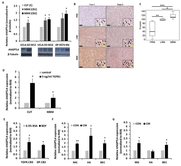
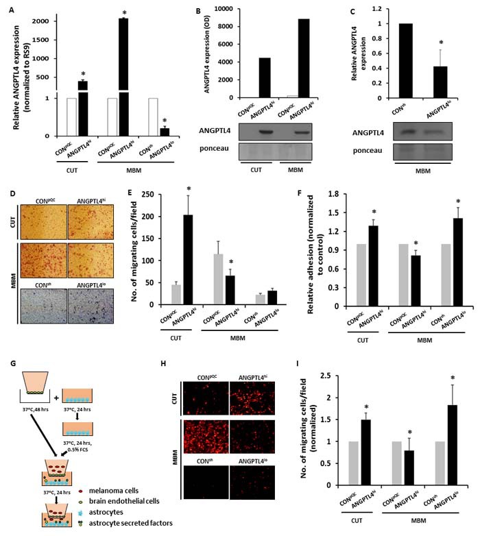
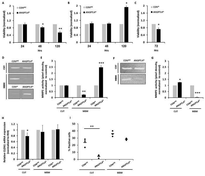
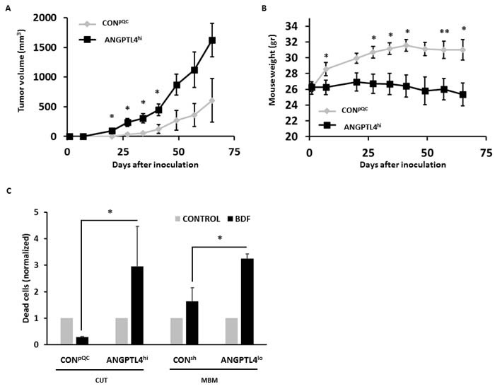
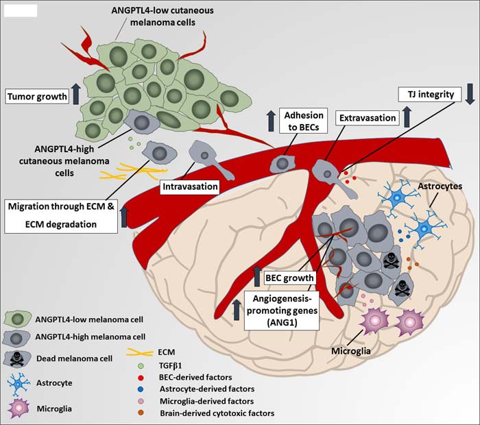
In an ongoing effort to identify molecular determinants regulating melanoma brain metastasis, we previously identified Angiopoietin-like 4 (ANGPTL4) as a component of the molecular signature of such metastases. The aim of this study was to determine the functional significance of ANGPTL4 in the shaping of melanoma malignancy phenotype, especially in the establishment of brain metastasis. We confirmed that ANGPTL4 expression is significantly higher in cells metastasizing to the brain than in cells from the cutaneous (local) tumor from the same melanoma in a nude mouse xenograft model, and also in paired clinical specimens of melanoma metastases than in primary melanomas from the same patients. In vitro experiments indicated that brain-derived soluble factors and transforming growth factor β1 (TGFβ1) up-regulated ANGPTL4 expression by melanoma cells. Forced over-expression of ANGPTL4 in cutaneous melanoma cells promoted their ability to adhere and transmigrate brain endothelial cells. Over-expressing ANGPTL4 in cells derived from brain metastases resulted in the opposite effects. In vivo data indicated that forced overexpression of ANGPTL4 promoted the tumorigenicity of cutaneous melanoma cells but did not increase their ability to form brain metastasis. This finding can be explained by inhibitory activities of brain-derived soluble factors. Taken together these findings indicate that ANGPTL4 promotes the malignancy phenotype of primary melanomas of risk to metastasize to the brain.

Keywords: ANGPTL4; TGFβ1; brain; melanoma; metastasis.


Conflict of interest statement

CONFLICTS OF INTEREST The Authors do not have any conflicts of interest.

Figures

Figure 1
Figure 1. ANGPTL4 expression during melanoma progression to brain metastasis
A. ANGPTL4 protein expression level in UCLA-SO-M12, UCLA-SO-M16 and DP-0574-Me cutaneous (CUT) and melanoma brain metastasizing (MBM) variants of first and second IC inoculation cycle was analyzed using Western blotting. The obtained values were normalized to β-Tubulin. The bars represent the relative expression of ANGPTL4 (normalized to RS9), compared to control, untreated cells + SD obtained in one measurement in at least three independent experiments. *P < 0.05. B., C. ANGPTL4 expression in paired samples of primary melanoma (PRM), melanoma lymph node metastasis (LNM), and melanoma brain metastasis (MBM) derived from melanoma patients. (B) Representative IHC staining with anti-ANGPTL4 Ab for PRM, LNM and MBM specimens. Black bars indicate 100µm. The insets show a magnification of the melanoma lesions. Black arrowheads indicate ANGPTL4 positive melanoma cells. Yellow bars indicate 20µm. (C) Box plot comparing H score for PRM, LNM and MBM. *P < 0.01, **P < 0.005, ***P < 0.0005. D. Melanoma cells were incubated with 5ng/ml TGFβ1 for 4 hrs. Following stimulation, RT-qPCR analysis was performed to determine the mRNA expression level of ANGPTL4. The bars represent the relative expression of ANGPTL4 (normalized to RS9), compared to control, untreated cells + SD obtained in one measurement in at least three independent experiments. *P < 0.05. E. Brains of BALB/c mice were harvested, and BDF were prepared after 24 hrs (see Materials and Methods) and added to melanoma cells for 24 hrs at 37°C. Melanoma cells treated with 0.5% BSA supplemented RPMI-1640 served as control. Following stimulation, RT-qPCR analysis was performed to determine the mRNA expression level of ANGPTL4. The bars represent the relative expression of ANGPTL4 (normalized to RS9), compared to control, untreated cells + SEM obtained in one measurement in at least three independent experiments. *P < 0.05. F., G. CM of microglia (MG), astrocytes (HA) and BEC was collected, and added to YDFR.CB3 (F) and DP.CB2 (G) melanoma cells for 24 hrs at 37°C. Melanoma cells treated with starvation medium served as control. Following stimulation, RT-qPCR analysis was performed to determine the mRNA expression level of ANGPTL4. The bars represent the relative expression of ANGPTL4 (normalized to RS9), compared to control, untreated cells + SEM obtained in one measurement in at least three independent experiments. *P < 0.05, **P < 0.005.
Figure 2
Figure 2. ANGPTL4 controls the malignancy phenotype of cutaneous and brain metastasizing melanoma variants
A.-C. Cutaneous (CUT) and melanoma brain metastasizing (MBM) variants were transduced with an ANGPTL4 cDNA-containing construct (ANGPTL4hi) or with the backbone construct pQCXIP (CONpQC). MBM cells were transduced with a mixture of 4 different shANGPTL4-containing constructs (ANGPTL4lo), or with the control construct (CONsh). The efficacy of ANGPTL4 over-expression or down-regulation was verified: (A) RT-qPCR analysis was performed to determine the mRNA expression level of ANGPTL4. The bars represent the relative ANGPTL4 expression (normalized to RS9) in ANGPTL4hi or ANGPTL4lo cells compared to control cells + SEM obtained in one measurement in at least three independent experiments. *P < 0.05. (B, C) Supernatants of the transduced cells were subjected to Western blot analysis. ANGPTL4 was detected by specific Abs. The bars represent the relative ANGPTL4 expression in ANGPTL4hi or ANGPTL4lo cells compared to control cells + SEM obtained in one measurement in two-three independent experiments. *P < 0.05. Representative blots are shown. Ponceau staining was used for loading control. D., E. Melanoma cells were allowed to migrate through collagen coated transwells for 24 hrs. The migrated cells were fixed and counted. (D) Representative images are presented (X10 magnification). (E) The bars represent the average number of migrating cells per field in three independent experiments performed in triplicates + SEM. *P < 0.05. F. BEC were cultured for 24 hrs to form a confluent monolayer and stimulated with 100 units/ml TNFα and IFNγ for additional 24 hrs. mCherry- or GFP-expressing melanoma cells were added and allowed to adhere for 30 min at 37°C. The fluorescence signal of labeled cells was measured before and after removal of non-adherent cells. The bars represent the average % adherent cells (normalized to control cells) + SEM in at least three independent experiments. Six replicates were performed in each experiment. *P < 0.05. G.-I. BEC were cultured for 48 hrs on the upper side of the apical chamber of transwell inserts to form a confluent monolayer. Astrocytes were cultured in 24-well plates for 24 hrs and starved for additional 24 hrs to allow the secretion of soluble factors. mCherry-expressing melanoma cells were added onto the BEC monolayer and allowed to migrate for 24 hrs towards the astrocytes (G). The migrated cells were fixed and counted. (H) Representative images are presented (X10 magnification). (I) The bars represent the average number of migrating cells per field (normalized to control cells) in at least three independent experiments performed in triplicates + SEM. *P < 0.05.
Figure 3
Figure 3. Secretion of bioactive factors from melanoma cells is dependent on ANGPTL4 expression level
A.-C. Melanoma cells were cultured for 24 hrs, then starved for additional 24 hrs, to allow secretion of melanoma-soluble factors. CM was collected and added to BEC. Cell viability of BEC grown with CM of cutaneous CONpQC and ANGPTL4hi melanoma cells (A) or MBM CONpQC and ANGPTL4hi melanoma cells (B) was monitored after 24, 48 and 120 hrs. Cell viability of BEC grown with CM of MBM CONsh and ANGPTL4lo melanoma cells (C) was monitored after 72 hrs. Viability was determined using XTT-based assay. Absorbance at 450 nm was determined for each well, and subtraction of nonspecific readings (measured at 630 nm) was calculated. The bars represent the average viability of the BEC grown with ANGPTL4hi or ANGPTL4lo melanoma CM relative to those grown in control melanoma CM + SD in three independent experiments. Six replicates were performed in each experiment. *P < 0.05, **P < 0.05. D.-G. Melanoma cells were seeded for 24 hrs and then cultured in serum-free media for additional 24 hrs. CM were collected and subjected to gelatin and collagen zymography. (D, F) Images of one representative experiment out of three are presented. A ~66 kDa band indicating the active form of MMP-2 was observed. (E, G) The bars represent the average MMP-2 activity (pixel density) as measured by densitometry, normalized to control cells + SD in one measurement in three independent experiments. *P < 0.05, **P < 0.0.1, ***P < 0.005. H., I. BECs were treated with melanoma CM for 24 hours. Then, CLDN1 expression was analyzed using RT-qPCR and flow cytometry. (H) The bars represent the relative CLDN1 mRNA expression (normalized to RS9) compared to control cells + SD obtained in one measurement in least three independent experiments. *P < 0.05. (I) BEC were trypsinized and analyzed for CLDN1 expression using flow cytometry. Dot plot histogram comparing % CLDN1 positive BEC. Each dot represents the value obtained in a single independent experiment. The line represents the average value in each variant. **P < 0.01.
Figure 4
Figure 4. ANGPTL4 alters the tumorigenic potential of melanoma cells
A. Volume of cutaneous tumors following subdermal implantation of CONpQC vs. ANGPTL4hi cutaneous cells. Tumor dimensions were measured using a caliper and volume was obtained as described in Materials and Methods. The average tumor volume + SEM is presented. *P < 0.05. B. Mice were weighed weekly following melanoma inoculation. The averege mouse weight + SD is presented. *P < 0.05. C. Melanoma cells were treated for 72 hrs with mouse BDF. Then, cells were trypsinized and cell death was determined by measuring DAPI incorporation. The bars represent the relative cell death of BDF treated cells compared to their controls + SEM obtained in one measurement in three independent experiments. *P < 0.05.
Figure 5
Figure 5. Proteomic expression analysis of ANGPTL4 high and low expressing cells
The expression of 305 proteins was examined using RPPA analysis in the ANGPTL4hi and ANGPTL4lo cells vs. their corresponding control cells. A., B., C. Comparisons were done for each pair of cells: (A) cutaneous ANGPTL4hi vs. CONpQC cells, (B) brain metastasizing ANGPTL4hi vs. CONpQC cells and (C) brain metastasizing ANGPTL4lo vs. CONsh cells. The tables show the normalized expression of differentially expressed proteins with FC < -1.25 or FC>1.25. D., E. Venn diagrams were used to compare lists of differentially expressed proteins. (D) Proteins differentially expressed in both comparisons obtained in (A) and (B). (E) Proteins differentially expressed in both comparisons obtained in (B) and (C). F. Protein-protein interactions of differentially expressed proteins with FC < -1.5 or FC>1.5 from all three comparisons. Circled in blue are proteins from comparison (A), circled in orange are proteins from comparison (B), and circled in green are proteins from comparison (C).
Figure 6
Figure 6. A proposed mechanism for ANGPTL4-mediated melanoma malignancy progression
A soluble factor in the microenvironment of the primary tumor transforming growth factor β1 (TGFβ1) induces the expression of ANGPTL4 in primary melanoma tumor cells. ANGPTL4 enhances their ability to migrate through extracellular matrix (ECM) components and to adhere and invade brain vasculature, for example by down-regulating the expression of cell-cell adhesion tight junction (TJ) molecules such as claudin-1 (CLDN1). Once arriving the brain, brain-derived soluble factors secreted by microglia, brain endothelial cells (BEC) and astrocytes, induce ANGPTL4 expression by brain metastasizing cells, what contributes to different phenotypes, such as resistance against brain-derived cytotoxic factors, enhancement of BEC growth and induction of angiogenesis-related genes such as angiopoietin 1 (ANG1) (data not shown) in BEC subjected to factors released from brain metastasizing cells, expressing high ANGPTL4 levels.

Similar articles

Cited by

References

    1. Marzese DM, Witz IP, Kelly DF, Hoon DS. Epigenomic landscape of melanoma progression to brain metastasis: unexplored therapeutic alternatives. Epigenomics. 2015;7:1303–11. - PubMed
    1. Gupta G, Robertson AG, MacKie RM. Cerebral metastases of cutaneous melanoma. Br J Cancer. 1997;76:256–9. - PMC - PubMed
    1. Bafaloukos D, Gogas H. The treatment of brain metastases in melanoma patients. Cancer Treat Rev. 2004;30:515–20. - PubMed
    1. Yano S, Shinohara H, Herbst RS, Kuniyasu H, Bucana CD, Ellis LM, Davis DW, McConkey DJ, Fidler IJ. Expression of vascular endothelial growth factor is necessary but not sufficient for production and growth of brain metastasis. Cancer Res. 2000;60:4959–67. - PubMed
    1. Klein A, Schwartz H, Sagi-Assif O, Meshel T, Izraely S, Ben Menachem S, Bengaiev R, Ben-Shmuel A, Nahmias C, Couraud PO, Witz IP, Erez N. Astrocytes facilitate melanoma brain metastasis via secretion of IL-23. J Pathol. 2015;236:116–27. - PubMed

LinkOut - more resources