

Thrombospondin-1 promotes cell migration, invasion and lung metastasis of osteosarcoma through FAK dependent pathway
- PMID: 29100277
- PMCID: PMC5652671
- DOI: 10.18632/oncotarget.17427
Thrombospondin-1 promotes cell migration, invasion and lung metastasis of osteosarcoma through FAK dependent pathway
Abstract
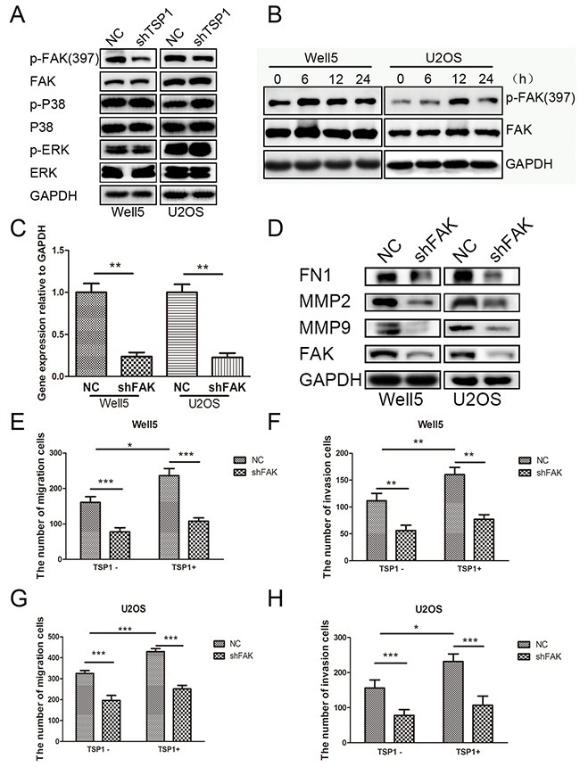
Microenvironment at the metastatic locus usually differs greatly from that present in the site of primary tumor formation and it has a significant impact on the fate of the extravasated cancer cells. We compared gene expression signatures of primary tumors and lung metastatic tumors, and identified Thrombospondin-1 (TSP1) as highly up-regulated in the lung metastatic tumors. Immunohistochemical staining further indicated that TSP1 protein expression was higher in lung metastatic tumors compared to primary tumors in both osteosarcoma xenograft model and human clinical samples. TSP1 mRNA level is significantly associated with the Enneking stage of osteosarcoma and lung metastasis. TGF-β pathways could stimulate the TSP1 expression in osteosarcoma cells. Knockdown of TSP1 expression in osteosarcoma cells dramatically suppressed cell wound healing, migration and invasion. Treatment with recombinant TSP1 protein in osteosarcoma cells significantly promoted cell wound healing, migration and invasion. Meanwhile, suppression of TSP1 in osteosarcoma cells resulted in decreased pulmonary metastasis in vivo. Mechanistically, TSP1 increased expression of metastasis related genes, including MMP2, MMP9 and Fibronectin 1. TSP1 promoted osteosarcoma cell motility through the activation of FAK pathway. Taken together, our study provides evidence of the contributions of TSP1 to the lung metastasis of osteosarcoma and suggests that this protein may represent a potential therapeutic target for osteosarcoma lung metastasis.
Keywords: FAK; lung metastasis; osoteosarcoma; thrombospondin-1.
Conflict of interest statement
CONFLICTS OF INTEREST We have no potential conflicts of interest to declare.
Figures


Similar articles
-
Spondin 1 promotes metastatic progression through Fak and Src dependent pathway in human osteosarcoma.Biochem Biophys Res Commun. 2015 Aug 14;464(1):45-50. doi: 10.1016/j.bbrc.2015.05.092. Epub 2015 May 29. Biochem Biophys Res Commun. 2015. PMID: 26032498
-
Thrombospondin-1 Silencing Improves Lymphocyte Infiltration in Tumors and Response to Anti-PD-1 in Triple-Negative Breast Cancer.Cancers (Basel). 2021 Aug 12;13(16):4059. doi: 10.3390/cancers13164059. Cancers (Basel). 2021. PMID: 34439212 Free PMC article.
-
BMPR2 promotes invasion and metastasis via the RhoA-ROCK-LIMK2 pathway in human osteosarcoma cells.Oncotarget. 2017 Apr 24;8(35):58625-58641. doi: 10.18632/oncotarget.17382. eCollection 2017 Aug 29. Oncotarget. 2017. PMID: 28938584 Free PMC article.
-
Regulation of tumor growth and metastasis by thrombospondin-1.FASEB J. 1996 Aug;10(10):1183-91. FASEB J. 1996. PMID: 8751720 Review.
-
How the NOTCH pathway contributes to the ability of osteosarcoma cells to metastasize.Cancer Treat Res. 2009;152:479-96. doi: 10.1007/978-1-4419-0284-9_28. Cancer Treat Res. 2009. PMID: 20213410 Review.
Cited by
-
The relationship between the Hippo signaling pathway and bone metastasis of breast cancer.Front Oncol. 2023 May 15;13:1188310. doi: 10.3389/fonc.2023.1188310. eCollection 2023. Front Oncol. 2023. PMID: 37256184 Free PMC article. Review.
-
A review: hippo signaling pathway promotes tumor invasion and metastasis by regulating target gene expression.J Cancer Res Clin Oncol. 2021 Jun;147(6):1569-1585. doi: 10.1007/s00432-021-03604-8. Epub 2021 Apr 17. J Cancer Res Clin Oncol. 2021. PMID: 33864521 Free PMC article. Review.
-
Stromal cartilage oligomeric matrix protein as a tumorigenic driver in ovarian cancer via Notch3 signaling and epithelial-to-mesenchymal transition.J Transl Med. 2024 Apr 13;22(1):351. doi: 10.1186/s12967-024-05083-0. J Transl Med. 2024. PMID: 38615020 Free PMC article.
-
Deep learning untangles the resistance mechanism of p53 reactivator in lung cancer cells.iScience. 2023 Nov 1;26(12):108377. doi: 10.1016/j.isci.2023.108377. eCollection 2023 Dec 15. iScience. 2023. PMID: 38034356 Free PMC article.
-
Thrombospondin-1 induced programmed death-ligand 1-mediated immunosuppression by activating the STAT3 pathway in osteosarcoma.Cancer Sci. 2022 Feb;113(2):432-445. doi: 10.1111/cas.15237. Epub 2021 Dec 23. Cancer Sci. 2022. PMID: 34927311 Free PMC article.
References
-
- Liu Y, Cao X. Characteristics and Significance of the Pre-metastatic Niche. Cancer Cell. 2016;30:668–81. - PubMed
LinkOut - more resources
Full Text Sources
Other Literature Sources
Miscellaneous
