. 2017 Jul 12;8(44):76153-76164.
doi: 10.18632/oncotarget.19190. eCollection 2017 Sep 29.

Long non-coding RNA MIAT promotes breast cancer progression and functions as ceRNA to regulate DUSP7 expression by sponging miR-155-5p

Affiliations

Long non-coding RNA MIAT promotes breast cancer progression and functions as ceRNA to regulate DUSP7 expression by sponging miR-155-5p

Tian Luan et al. Oncotarget. .

Abstract

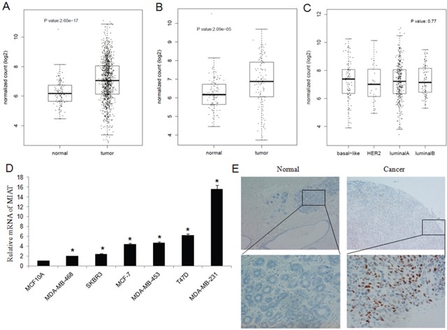
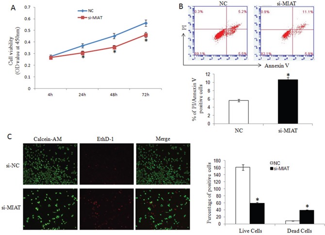
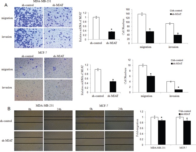
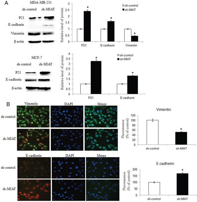
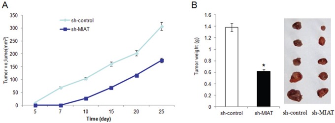
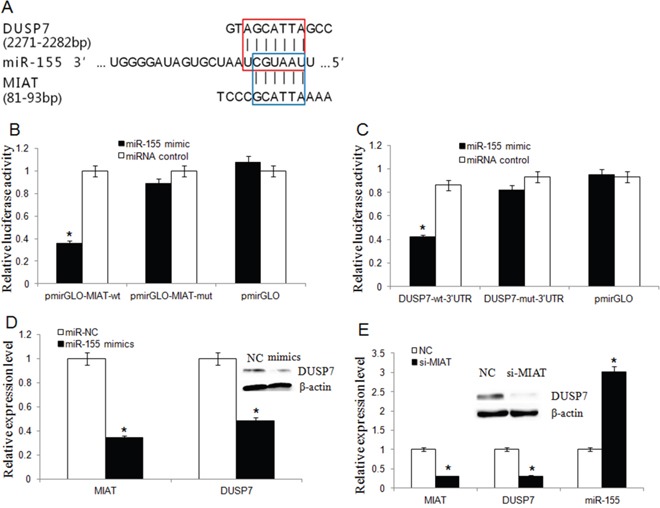
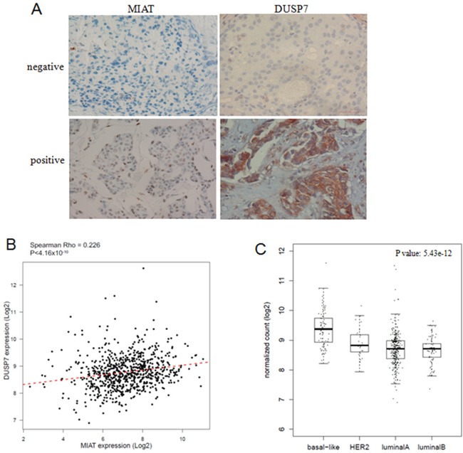
Long non-coding RNAs (lncRNA) have been reported as key regulators in the progression and metastasis of breast cancer. In this study, we found that the lncRNA myocardial infarction associated transcript (MIAT) expression was upregulated in breast cancer in The Cancer Genome Atlas (TCGA) data sets. We validated that MIAT was higher in breast cancer cell lines and advanced breast tumors than in normal controls. And MIAT overexpression associated with TNM stage and lymphnode metastasis. Knockdown MIAT inhibited breast cancer cell proliferation and promoted apoptosis. Also MIAT downregulation suppressed epithelial-mesenchymal transition (EMT) and decreased migration and invasion in MDA-MB-231 and MCF-7 breast cancer cell lines. More importantly, knockdown MIAT inhibited tumor growth in vivo. Our results suggested that MIAT acted as a competing endogenous RNA (ceRNA) to regulate the expression of dual specificity phosphatase 7 (DUSP7) by taking up miR-155-5p in breast cancer. There were positive correlation between MIAT and DUSP7 expression in breast cancer patients. We conclude that MIAT promotes breast cancer progression and functions as ceRNA to regulate DUSP7 expression by sponging miR-155-5p in breast cancer.

Keywords: DUSP7; MIAT; breast cancer; ceRNA; miR-155-5p.


Conflict of interest statement

CONFLICTS OF INTEREST The authors declare no competing financial interests.

Figures

Figure 1
Figure 1. Expression of MIAT in TCGA, breast cancer cell lines and tissues
(A) MIAT expression was higher in breast cancer tissues than in normal breast tissues from TCGA dataset (P<0.05). (B) MIAT expression was higher in breast cancer tissues than in paired normal breast tissues from TCGA dataset (P<0.05). (C) There were no significance of MIAT expression among different breast cancer subtypes from TCGA dataset (P=0.77). (D) MIAT expression was higher in breast cancer cell lines than in normal breast epithelial cell line MCF-10A. *P <0.05, Two-side Student's t-test; n = 3. (E) RNA Scope® technology detection showed that MIAT RNA was significant overexpression in human breast cancer tissues than in adjacent normal tissues (up ×100; bottom ×400).
Figure 2
Figure 2. MIAT downregulation inhibited breast cancer cell proliferation and promoted apoptosis
(A) CCK-8 assays were performed to measure the proliferation of MDA-MB-231 cells. *P< 0.05, Two-side Student's t-test; n=3. (B) MIAT downregulation promoted apoptosis as shown by increased PI and Annexin V positive cells. *P <0.05, Two-side Student's t-test; n = 3. (C) Cell apoptosis was analyzed using Calcein-AM/EthD-1 staining. Green: live cells, Red: dead or dying cells (×100). *P <0.05, Two-side Student's t-test; n = 3.
Figure 3
Figure 3. MIAT downregulation inhibited migration and invasion of MDA-MB-231 and MCF-7 breast cancer cells
(A) Transwell migration and invasion assays were used to evaluate MDA-MB-231 and MCF-7 cell numbers with or without MIAT downregulation. Cells were stained with crystal violet after cultured 24h and counted for statistical analysis (×200). The qRT-PCR was done to verify the MIAT expression level. *P <0.05, Two-side Student's t-test; n = 3. (B) The scratch-wound gap of MDA-MB-231 and MCF-7 cells with or without MIAT downregulation was photographed before and after 24 h incubation. After performing the wound healing test, the results were analyzed by measuring the range of migrating cells from 3 different fields for each wound (×50). *P <0.05, Two-side Student's t-test.
Figure 4
Figure 4. MIAT downregulation inhibited breast cancer cell EMT
(A) Western blot analysis showed that knockdown of MIAT increased P21 and E-cadherin expression both in MDA-MB-231 and MCF-7 cells. The vimentin level was inhibited in MDA-MB-231 cell after MIAT knockdown but no vimentin was found in MCF-7 cell (data not shown). β-actin was detected as the loading control. *P <0.05, Two-side Student's t-test; n = 3. (B) Immunofluoresence assay was conducted to detect vimentin and E-cadherin expression in MDA-MB-231 cells. The expression of vimentin was lower but E-cadherin was higher in cells transfected with sh-MIAT than in sh-control cells. Nuclei, blue; vimentin (up) or E-cadherin (bottom), green; Cells transfected with lentivirus, red (×400). Results were presented as a relative percentage to control (defined as 100%). *P <0.05, Two-side Student's t-test; n = 3.
Figure 5
Figure 5. Knockdown of MIAT inhibited MDA-MB-231 cells growth in vivo
(A). Summary of tumor volume of nude mice which were measured in every 5 days in sh-control and sh-MIAT group, respectively. (B) Tumor weight and representation picture of tumor formation of xenograft in nude mice in sh-control and sh-MIAT group, respectively (each group n=5).
Figure 6
Figure 6. MIAT and DUSP7 shared a common miR-155-5p binding site
(A) Bioinformatics analysis revealed that MIAT and DUSP7 shared a common miR-155-5p binding site. The red box represented the binding site for miR-155-5p on the DUSP7 mRNA 3′-UTR, and the blue box represented the binding site for miR-155-5p on MIAT. (B) Luciferase activity in HEK293T cells cotransfected with miR-155-5p mimics and luciferase reporters containing control vector, pmir-Glo-MIAT-wt and pmir-Glo-MIAT-mut (*P<0.05, Two-side Student's t-test; n = 3). miR-155-5p mimics reduced the luciferase activity of wt MIAT reporter vector but not that of mut MIAT reporter vector. (C) MiR-155-5p significantly inhibited the luciferase activity of the DUSP7 wt 3′-UTR but not that of the mutant (*P <0.05, Two-side Student's t-test; n = 3). (D) MiR-155-5p overexpression downregulated the endogenic MIAT and DUSP7 expression. (*P <0.05, Two-side Student's t-test; n = 3). Western blot assay showed that miR-155-5p overexpression silenced DUSP7 protein expression. (E) The miR-155-5p level was increased but DUSP7 mRNA level was decreased in MDA-MB-231 cells after MIAT knockdown. (Data are presented as mean ± SD, n=3. *P <0.05). Western blot assay showed that knockdown of MIAT triggered a significant silencing effect on endogenous DUSP7 protein.
Figure 7
Figure 7. MIAT and DUSP7 had positive association
(A) Consecutive sections of MIAT and DUSP7 expression in breast cancer TMA. MIAT RNA was detected by RNA Scope 2.0® technology and DUSP7 protein was detected by IHC (×400). MIAT located at nucleus and DUSP7 located at cytoplasm. (B) The linear correlations between the expression level of MIAT and DUSP7 in breast cancer tissues from TCGA database (R=0.226, P<0.05). The data were obtained using the logistic regression analysis. (C) The DUSP7 expression was higher in basal-like breast cancer subtypes from TCGA database (P<0.05).

References

    1. Stagl JM, Bouchard LC, Lechner SC, Blomberg BB, Gudenkauf LM, Jutagir DR, Gluck S, Derhagopian RP, Carver CS, Antoni MH. Long-term psychological benefits of cognitive-behavioral stress management for women with breast cancer: 11-year follow-up of a randomized controlled trial. Cancer. 2015;121:1873–1881. - PMC - PubMed
    1. DeSantis C, Ma J, Bryan L, Jemal A. Breast cancer statistics, 2013. CA Cancer J Clin. 2014;64:52–62. - PubMed
    1. Batista PJ, Chang HY. Long noncoding RNAs: cellular address codes in development and disease. Cell. 2013;152:1298–1307. - PMC - PubMed
    1. Fatica A, Bozzoni I. Long non-coding RNAs: new players in cell differentiation and development. Nat Rev Genet. 2014;15:7–21. - PubMed
    1. Liu D, Yu X, Wang S, Dai E, Jiang L, Wang J, Yang Q, Yang F, Zhou S, Jiang W. The gain and loss of long noncoding RNA associated-competing endogenous RNAs in prostate cancer. Oncotarget. 2016;7:57228–57238. doi: 10.18632/oncotarget.11128. - DOI - PMC - PubMed