. 2017 Aug 10;8(44):76881-76897.
doi: 10.18632/oncotarget.20173. eCollection 2017 Sep 29.

PAX2 maintains the differentiation of mouse oviductal epithelium and inhibits the transition to a stem cell-like state

Affiliations

PAX2 maintains the differentiation of mouse oviductal epithelium and inhibits the transition to a stem cell-like state

Kholoud Alwosaibai et al. Oncotarget. .

Abstract

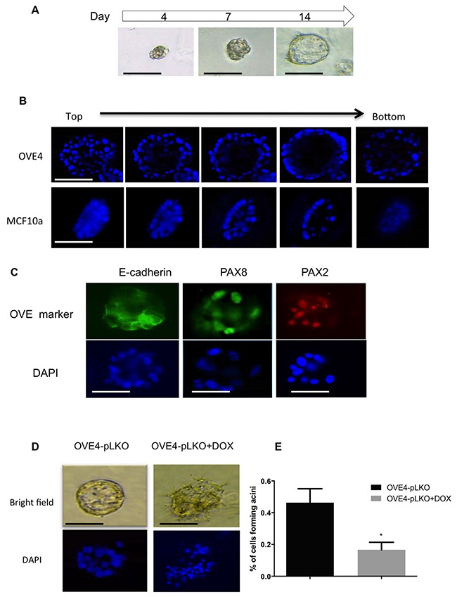
Recent studies have provided evidence that the secretory cells of the fallopian tube (oviduct) are a probable origin for high-grade serous ovarian carcinoma. In addition to secretory cells, the fallopian tube epithelium consists of ciliated cells and CD44+ undifferentiated stem-like cells. Loss of PAX2 expression is recognized as an early event in epithelial transformation, but the specific role of PAX2 in this transition is unknown. The aim of this study was to define the role of PAX2 in oviductal epithelial (OVE) cells and its response to transforming growth factor β1 (TGFβ), characterizing specifically its potential involvement in regulating stem cell-like behaviors that may contribute to formation of cancer-initiating cells. Treatment of primary cultures of mouse OVE cells with TGFβ induced an epithelial-mesenchymal transition (EMT) associated with decreased expression of PAX2 and an increase in the fraction of cells expressing CD44. PAX2 knockdown in OVE cells and overexpression in ovarian epithelial cells confirmed that PAX2 inhibits stem cell characteristics and regulates the degree of epithelial differentiation of OVE cells. These results suggest that loss of PAX2, as occurs in serous tubal intraepithelial carcinomas, may shift secretory cells to a more mesenchymal phenotype associated with stem-like features.

Keywords: PAX2; epithelial-mesenchymal transition; fallopian tube; ovarian cancer; stem cells.


Conflict of interest statement

CONFLICTS OF INTEREST The authors declare no conflicts of interest.

Figures

Figure 1
Figure 1. Characterization of clonal lines of oviductal epithelial cells
(A) OVE4, OVE16 and OVE22 cells express oviductal cell markers (Cdh1, Pax2, Pax8, Ovgp1 and FoxJ1). (B) TGFβ treatment (10ng/ml) for 7 days induced fibroblast-like phenotypes in OVE clonal cell lines and (C) inhibited cell proliferation in all OVE lines, as assessed after 7 days. Data presented in histograms are mean ± SEM. Scale bars indicate 100μm. All experiments were performed at least three times. In A, different letters denote values that are significantly different (p<0.05). In C, * indicates p<0.05; ** indicates p<0.01.
Figure 2
Figure 2. TGFβ induces EMT in oviductal epithelial cells
(A) TGFβ pre-treatment for 3 days increased cell migration of OVE4 and OVE22 cells, as seen 24 hours after wounding. (B) Western blot and densitometric analysis for epithelial and EMT proteins (E-cadherin, SNAIL and Vimentin) in OVE4 cells treated with TGFβ for up to 3 days. (C) qPCR analysis for mRNA encoding for epithelial and EMT genes for OVE4 cells treated for one day with TGFβ. Data are from three independent experiments and are presented in histograms as mean ± SEM. Scale bars indicate 400μm. * indicates p<0.05; ** indicates p<0.01.
Figure 3
Figure 3. TGFβ increases the expression of stem cell markers in oviductal epithelial cells
(A and B) OVE cells form spheres in low attachment plates and average sphere size is increased in OVE4 cells by TGFβ treatment. (C) Relative expression of mRNA encoding for stem cell markers in OVE4 shows that TGFβ treatment for 7 days significantly up-regulates CD44 and, to a lesser extent, Sca1 mRNA. (D) Western blots and densitometric analysis of those blots normalized to β-actin show increased expression of CD44 in OVE4 and OVE16 after 1 day of TGFβ treatment. (E) Sphere formation capacity of CD44 positive and negative populations sorted by flow cytometry. All data are from three independent experiments, Data presented in histograms are mean ± SEM. Scale bar in (A) is 100μm. * indicates p<0.05; ** p<0.01; *** p<0.001.
Figure 4
Figure 4. Immunohistochemistry shows CD44 staining only in the fimbriae and a few cells in the ovarian surface epithelium
Figure 5
Figure 5. TGFβ suppresses Pax2 expression in oviductal epithelial cells
(A) qPCR analysis for mRNA encoding Pax2 for OVE4 and OVE16 cells treated for one day with TGFβ. (B) Western blots and histogram of densitometric analysis show decreased abundance of PAX2 after 1-2 days of 10ng/ml TGFβ treatment of OVE4 and OVE16 cells. (C) Western blot shows time course of TGFβ treatment resulting in coincident reduction of PAX2 abundance with increased SMAD2 phosphorylation. All data are representative of three independent experiments except SMAD2 western blot that is representative of two independent experiments. Data presented in histograms are mean ± SEM. * p<0.05; ** p<0.01; *** p<0.001.
Figure 6
Figure 6. PAX2 down-regulates CD44 expression
(A) qPCR analysis shows decreased levels of Pax2 mRNA is associated with increased levels of CD44 mRNA in OVE4 and OVE16 cells after 1 day of TGFβ treatment. (B) Western blot and densitometric analysis show PAX2 knockdown after doxycycline treatment of OVE cells transduced with Pax2 shRNA constructs. (C) Knockdown of PAX2 increases mRNA for stem cell markers CD44, Sca-1 and CD133. (D and E) Sphere-forming capacity of OVE cells increased after PAX2 knockdown only in OVE4 cells, and there was no effect on sphere size. All experiments were performed at least three times and for experiments shown in B-E, samples were collected for analysis 4-6 cell passages after initiating doxycycline treatment. * p<0.05; ** p<0.01; *** p<0.001.
Figure 7
Figure 7. PAX2 decreases stem cell characteristics in mouse OSE cells (M1102)
(A) Western blot shows the forced expression of PAX2 in M1102, with subsequent loss of expression in cells treated with adenovirus expressing Cre recombinase (AdCre) to delete the floxed PAX2 construct. (B-D) Overexpression of PAX2 in M1102 cells decreases sphere formation in suspension (N=3), but has no effect on sphere size. (E) Representative western blot shows that expression of PAX2 in M1102 cells decreases both variant and standard forms of CD44. (F) Densitometric analysis of CD44v and CD44s protein levels in M1102 cells from western blots normalized to β-actin (N=3). (G) Immunofluorescence staining shows that induction of Pax2 expression in M1102 cells decreases abundance of CD44. (H) qPCR analysis for stem cell markers shows that expression of Pax2 decreases mRNA encoding for Sca-1 and Lgr5 in M1102 cells (N=3). Data are presented as mean ± SEM. Scale bar in (B) indicates 100μm. Different letters denote values that are significantly different (p<0.05).
Figure 8
Figure 8. PAX2 is required for epithelial differentiation of OVE cells
(A) Formation of luminal structure by OVE cells cultured in matrigel starts with small spheres by day 4 and ends with spheroids with a lumen by day 14. (B) Immunofluorescence by Z-stack imaging starting from the top of spheroids formed by OVE4 and MCF10a cells in matrigel after 14 days of culture shows the lumen in the middle of each spheroid. (C) OVE4-derived luminal structures express E-cadherin and the oviductal markers, PAX2 and PAX8, as detected by immunofluorescence. (D) Knockdown of PAX2 (right panels) disrupts the formation of luminal structures by OVE4 cells (left panels), which is quantified in (E). Data are presented as mean ± SEM (N=3). Scale bars in (A-C) indicate 100μm. * indicates p<0.05.

Similar articles

Cited by

References

    1. Auersperg N, Wong AS, Choi KC, Kang SK, Leung PC. Ovarian surface epithelium: biology, endocrinology, and pathology. Endocr Rev. 2001;22:255–288. - PubMed
    1. Yao DS, Li L, Garson K, Vanderhyden BC. The mouse ovarian surface epithelium cells (MOSE) transformation induced by c-myc/K-ras. Zhonghua Zhong Liu Za Zhi. 2006;28:881–885. - PubMed
    1. Laviolette LA, Hodgkinson KM, Minhas N, Perez-Iratxeta C, Vanderhyden BC. 17beta-estradiol upregulates GREB1 and accelerates ovarian tumor progression in vivo. Int J Cancer. 2014;135:1072–1084. - PMC - PubMed
    1. Hodgkinson KM, Vanderhyden BC. Consideration of GREB1 as a potential therapeutic target for hormone-responsive or endocrine-resistant cancers. Expert Opin Ther Targets. 2014;18:1065–1076. - PubMed
    1. Kim J, Coffey DM, Ma L, Matzuk MM. The ovary is an alternative site of origin for high-grade serous ovarian cancer in mice. Endocrinology. 2015;156:1975–1981. - PMC - PubMed