. 2017 Aug 24;8(44):77317-77329.
doi: 10.18632/oncotarget.20469. eCollection 2017 Sep 29.

Targeting Hsp27/eIF4E interaction with phenazine compound: a promising alternative for castration-resistant prostate cancer treatment

Affiliations

Targeting Hsp27/eIF4E interaction with phenazine compound: a promising alternative for castration-resistant prostate cancer treatment

Hajer Ziouziou et al. Oncotarget. .

Erratum in

Abstract

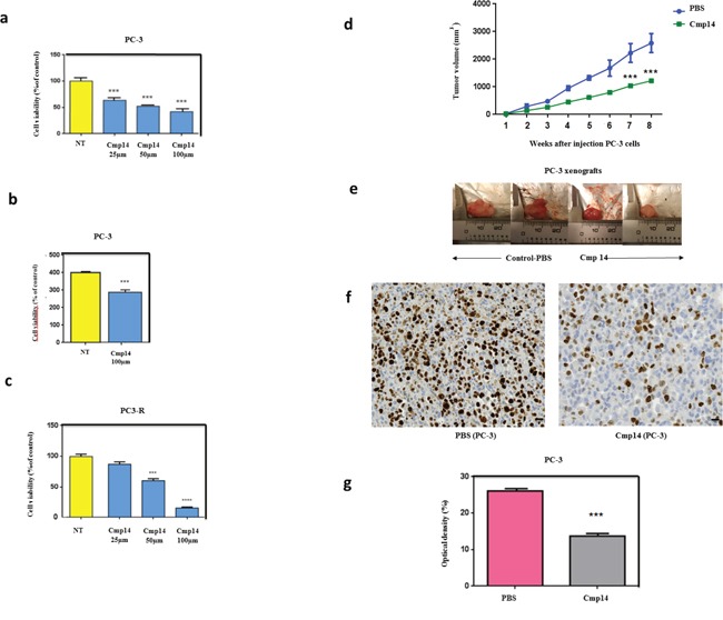
The actual strategy to improve current therapies in advanced prostate cancer involves targeting genes activated by androgen withdrawal, either to delay or prevent the emergence of the castration-refractory phenotype. However, these genes are often implicated in several physiological processes, and long-term inhibition of survival proteins might be accompanied with cytotoxic effects. To avoid this problem, an alternative therapeutic strategy relies on the identification and use of compounds that disrupt specific protein-protein interactions involved in androgen withdrawal. Specifically, the interaction of the chaperone protein Hsp27 with the initiation factor eIF4E leads to the protection of protein synthesis initiation process and enhances cell survival during cell stress induced by castration or chemotherapy. Thus, in this work we aimed at i) identifying the interaction site of the Hsp27/eIF4E complex and ii) interfere with the relevant protein/protein association mechanism involved in castration-resistant progression of prostate cancer. By a combination of experimental and modeling techniques, we proved that eIF4E interacts with the C-terminal part of Hsp27, preferentially when Hsp27 is phosphorylated. We also observed that the loss of this interaction increased cell chemo-and hormone-sensitivity. In order to find a potential inhibitor of Hsp27/eIF4E interaction, BRET assays in combination with molecular simulations identified the phenazine derivative 14 as the compound able to efficiently interfere with this protein/protein interaction, thereby inhibiting cell viability and increasing cell death in chemo- and castration-resistant prostate cancer models in vitro and in vivo.

Keywords: Hsp27/eIF4E interaction; prostate cancer; protein-protein interaction inhibition.


Conflict of interest statement

CONFLICTS OF INTEREST The authors declare no conflicts of interest.

Figures

Figure 1
Figure 1. Modeling of Hsp27 with eIF4E and phosphorylation status of Hsp27 reveal that Hsp27 C-terminal domain and phosphorylation are essential for protein binding and loss of this interaction increases cell chemo and hormone-sensitivity
(a) Schematic representation of Hsp27 wild type (WT) and truncated mutant forms of Hsp27 (N1, N2, and C1). Mutants N1 and N2 correspond to two different variants of the N-terminal region of Hsp27 protein (residues 1-93, and 1-173, respectively), whereas mutant C1 corresponds to the Hsp27 C-terminal region, containing the last 113 protein residues (residues 93-205). The full-length WT Hsp27 sequence was used as control. No endogenous Hsp27 expression REG cells transiently transfected or not (NT) with plasmids containing WT Hsp27 and N1, N2 and C1 and proteins were extracted for: (b) Western Blot analysis of histidine and vinculin protein levels from total cell lysates (TCL) and (c) Western Blot analysis of histidine and eIF4E protein levels after immunoprecipitation (IP) using anti-eIF4E antibody (d) MTT quantification of REG cells viability. (e) Schematic representation of Hsp27 wild type (WT) and phosphorylation mutants of Hsp27 (3A and 3D) used in this study. In the former case, the 3A mutant of Hsp27 was specifically constructed by replacing the serine residues 15, 78 and 82 with alanine; concomitantly, the 3D Hsp27 isoforms was obtained by replacing the same serine residues with aspartic acid. (f) Western Blot analysis of histidine and vinculin protein levels from total cell lysates (TCL) and (g) Western Blot analysis of histidine and eIF4E protein levels after immunoprecipitation (IP) using anti-eIF4E antibody. (h) MTT quantification of REG cells viability was performed on cells transiently transfected with plasmids containing WT Hsp27 and phosphorylation mutants of Hsp27 prior to treatment with docetaxel in serum-free media (a condition mimicking androgen deprivation) *** P≤0.001. (i) Overall view of an equilibrated MD snapshot of the Hsp27 WT/eIF4E complex. The proteins are visualized by their van der Waals surfaces, colored as follow: Hsp27 WT, firebrick; eIF4E, khaki. The amino acids of Hsp27 mainly involved in binding eIF4E are highlighted as follows: purple, residues belonging to the WDPF domain; green, residues belonging to the flexible domain. See text for details. (j) Per residue enthalpic contribution to WT Hsp27 binding with eIF4E. Only those Hsp27 amino acids affording a meaningful contribution to protein-protein formation contributing (ΔHbind,res < -0.80 kcal/mol) are shown for clarity. From this analysis, it further appears that the Hsp27 α-crystallin domain is practically ineffective in Hsp27/eIF4E binding since residues belonging to this Hsp27 region display a negligible contribution to protein-protein binding enthalpy. (k) Overall view of equilibrated MD snapshots of the eIF4E in complex with Hsp27 N1, N2 and C1 truncated mutant isoforms. In each panel, the proteins are visualized by their van der Waals surfaces, colored as follows: Hsp27s, firebrick; eIF4E, khaki. The amino acids of Hsp27 mainly involved in binding eIF4E are highlighted as follows: purple, residues belonging to the WDPF domain; green, residues belonging to the flexible domain. Interestingly, the overall structure of Hsp27 N1 is de facto strongly affected by the loss of the α-crystallin domain, which plays a major role in the overall correct chaperon folding required for effective binding to eIF4E.
Figure 2
Figure 2. Validation of Hsp27/eIF4E interaction by BRET in whole living cells and in cells extracts and revelation of the chemical compound 14 as a specific inhibitor Hsp27/eIF4E by BRET screening
(a) To by-pass technical parameters that could prevent us from measuring a BRET signal (presence and/or position of the reporter proteins that could disrupt the Hsp27/eIF4E interaction, physical distance between Rluc and YFP), the interaction between these two proteins with all possible couple combinations (n=8) of plasmids were tested. These couples of plasmids were transfected separately in HEK293T cells at different concentrations in order to find the ideal ratios and obtain the BRET signal. For the construction of BRET plasmids, we merged each gene (Hsp27 and eIF4E) with Rluc or YFP in N-terminal (N-ter) or C-terminal (C-ter) part. After, we tested the interaction between these two proteins with all the possible couple combinations of plasmids, on BRET on living cells or cells extracts. (b) Hsp27/eIF4E interaction in BRET experiment was investigated in HEK293T cell extracts. The principle was similar to BRET assay in living cells; except that the plasmids were transfected separately in HEK293T cells and that the couple combinations were tested in vitro by mixing proteins extracted from these transfected cells. HEK293T cells were co-transfected with 0.2 μg of BRET donor plasmid N-terluc/eIF4E, and 0 to 1 μg of BRET acceptor plasmid Hsp27/YFPC-ter. The empty vector (pEYFP-C) was used to equalize DNA amounts to 1.2 μg in each sample. The reading of optic density was performed after the addition of coelenterazine in order to obtain the BRET signal. (c) HEK293T cells were transfected separately with a BRET donor plasmid N-terluc/eIF4E or BRET acceptor plasmid Hsp27/YFPC-ter. Δ=(YFP/Luc-YFP0/Luc0)*1000. (d) Derivatives of phenazines that have been described to have an anti-tumor activity as well as a structure similar to inhibitors of eIF4E/eIF4G interaction. (e) Compound 4E2RCat, which is described in literature to be an inhibitor of the eIF4E/eIF4G interaction. (f) HEK293T cells were transfected separately with a BRET donor plasmid N-terluc/eIF4E or BRET acceptor plasmid Hsp27/YFPC-ter. Total proteins were extracted from cells and used for BRET assay: 1 μg of lysate containing a BRET donor and 0 to 15 μg of lysate containing a BRET acceptor were pre-incubated separately with different concentrations (0, 20, 50 and 100μM; green, yellow, orange and red lines respectively) of compound 14 during 2h. As control experiment, cell extracts were pre-incubated with DMSO alone, at the higher concentration (1%) we used to dilute compound (control DMSO, blue line). Donor and acceptor were mixed for 30 min and the reading of optic density was performed after the addition of coelenterazine in order to obtain the BRET signal. (g) The same experiment was performed with another couple of protein: CCND3/luc and CDK6/YFP (4μg of lysate containing BRET donor was used). Δ=(YFP/Luc-YFP0/Luc0)*1000. (h) PC-3 cells were treated at 100μM with compound 14 (right panel, Bar=10 μm) and DMSO (left panel, Bar = 20μm) as control. Auto-fluorescence of compound 14 (green) and staining of the nucleus by DAPI (blue) was observed. PC-3 cells were treated with DMSO (control) or compound 14 during 48h and proteins were extracted for: (i) Western Blot analysis of Hsp27, eIF4E protein levels after immunoprecipitation (IP) using eIF4E rabbit antibody or IgG rabbit (control) (j) Western Blot analysis of Hsp27, eIF4E, Vinculin and protein levels from total protein extracts.
Figure 3
Figure 3. Molecular modeling and isothermal titration calorimetry (ITC) reveal the mechanism of Hsp27/eIF4E interaction inhibition by compound 14
(a) (Left panel) Zoomed view of an MD equilibrated snapshot of the 14 in complex with eIF4E. Specifically, the ligand is docked between a α-helix of eIF4E protein spanning residue L117*-E132* and a structurally hybrid region from E185* to I193*. The ligand is portrayed in ball-and-stick representation and colored by element (C, gray; N, blue; O, red). The main protein residues involved in compound binding are depicted as gold sticks and labelled. Transparent light blue spheres represent water oxygen atoms, while chlorine and sodium ions and counterions are shown as green and purple spheres, respectively. Hydrogen atoms are omitted for clarity. (Right Panel) Per residue binding enthalpy decomposition for eIF4E residues mainly involved in binding with 14. Only those Hsp27 amino acids affording a meaningful contribution to protein-drug formation (ΔHbind,res* < -0.50 kcal/mol) are shown for clarity. The network of stabilizing hydrophobic interaction involves the two C12 alkyl chains of the 14 and the side chain of the protein residue L117*, N118*, F129*, L189*, P190*, K192*, and I193*. The two amine substituents of 14 are engaged in two persistent polar interactions with the carboxylic side chain of E132* and E185*. In addition, a further stabilizing interaction via a weak hydrogen bond between the side chain of R128* and a nitrogen atom of the phenazine ring is detected. (b) (Left panel) MD simulation distance between the charged side chains of E132* and R186* in the eIF4E alone (red line) and in the eIF4E/14 complex (salmon line). In the eIF4E free protein, the two α-helixes spanning residues Q122*-I138* and R173*-G188*, respectively, are persistently stabilized by the presence of permanent interaction points between the side chain of their residues. In particular, a strong polar interaction between the charged side chain of the amino acids E132* and R186* is detected. As shown, the Average Dynamics Length (ADL) of this interaction between the two involved atoms is 2.96 Å, and its persistence is verified along the entire MD run. Conversely, the same distance progressively increases during the first part of the simulations and finally settles around 9Å when the eIF4F is simulated in presence of the ligand. The reason for this behavior can be explained by that fact that E132* is engaged in a polar interaction with 14, as demonstrated by the complementary trend exhibited of the corresponding distance between the carboxylic moiety of E132* and the amine group of 14 shown in the right panel (MD simulation distance between the N2 nitrogen atom of 14 and the charged side chain of E132* (blue line), and between the N3 nitrogen atom of 14 and the charged side chain of E185* (green line). (c) (Left panel) Overall view of an equilibrated MD snapshot of the Hsp27 WT/eIF4E/14 complex. All molecules are visualized by their van der Waals surfaces, colored as follow: WT Hsp27, firebrick; eIF4E, khaki; 14, navy blue. The amino acids of Hsp27 mainly involved in binding eIF4E are highlighted as follows: purple, residues belonging to the WDPF domain; green, residues belonging to the flexible domain. The residues of eIF4E mainly involved in binding with 14 are depicted in gold. (Upper right panel) Binding free energies (ΔGbind) and binding free energy differences (ΔGbind) for the WT Hsp27/eIF4E (plain filled columns) and the WT Hsp27/eIF4E/14 (patterned filled columns) complexes. (Lower right panel) Comparison of the clustered per residue enthalpic contribution to binding for WT Hsp27/eIF4E and WT Hsp27/eIF4E/14 complexes. (d) ITC experiment of 14/eIF4E binding: (left) raw data; (middle) titration curve; (right) binding thermodynamics parameters.
Figure 4
Figure 4. Compound 14 inhibits cell viability and increases apoptosis in vitro and in vivo
Cells viability using MTT assay (a) was performed on PC-3 non treated (NT) or treated cells with compound 14 (a) at different concentrations (25, 50, 100μM) during 48h. Cell death quantification (SubG0 phase) using flow cytometry (b) was performed on PC-3 cells non treated (NT) or treated with compound 14 at 100μM during 48h. Cell viability using MTT assay was also performed on PC-3-docetaxel resistant cells non treated (NT) or treated with compound 14 (c) at different concentrations (25, 50, 100μM) during 48h. (d) PC-3 cells were subcutaneously implanted in Node Scids by injection of 10 ×10 6 cells in the right flank of animals. When tumors reached 100mm3, mice were randomized in two groups that received twice a week an intra-peritoneal injection of PBS (control n = 6, blue) and phenazine 14 (n = 8, green) (1 mg/kg) for 8 weeks. Tumor volume was measured once weekly and calculated by the formula length x width x depth x 0.5236. Compound 14 reduced significantly PC-3 tumor volume by up to 50%. During the entire treatment period, all mice treated with PBS and 14 did not show any abnormal behavior, and no significant alteration of mice body weight was observed. (e) Photographs of PC-3 harvested tumors from mice that received i.p. compound 14 or control-PBS after an 8-week treatment (f) Ki-67 IHC staining of tumor tissues to assess tumor cells proliferation. (g) Distribution of tissue Ki-67 immunostaining intensity (measured as average optical density) according to the tumor treated with PBS and Compoud#14. Error bars represent the SE, **, P ≤0.01 and ***, P ≤ 0.001 by Statview software.

References

    1. Resnick MJ, Lacchetti C, Penson DF. Prostate cancer survivorship care guidelines: American Society of Clinical Oncology practice guideline endorsement. J Oncol Pract. 2015;11:e445–449. - PubMed
    1. Badrising S, van der Noort V, van Oort IM, van den Berg HP, Los M, Hamberg P, Coenen JL, van den Eertwegh AJ, de Jong IJ, Kerver ED, van Tinteren H, Bergman AM. Clinical activity and tolerability of enzalutamide (MDV3100) in patients with metastatic, castration-resistant prostate cancer who progress after docetaxel and abiraterone treatment. Cancer. 2014;120:968–975. - PubMed
    1. Katsogiannou M, Ziouziou H, Karaki S, Andrieu C, Henry de Villeneuve M, Rocchi P. The hallmarks of castration-resistant prostate cancers. Cancer Treat Rev. 2015;41:588–597. - PubMed
    1. Mymrikov EV, Seit-Nebi AS, Gusev NB. Large potentials of small heat shock proteins. Physiol Rev. 2011;91:1123–1159. - PubMed
    1. Acunzo J, Andrieu C, Baylot V, So A, Rocchi P. Hsp27 as a therapeutic target in cancers. Curr Drug Targets. 2014;15:423–431. - PubMed