

Tandem mass spectrometry assay of β-glucocerebrosidase activity in dried blood spots eliminates false positives detected in fluorescence assay
- PMID: 29100779
- PMCID: PMC5808899
- DOI: 10.1016/j.ymgme.2017.10.011
Tandem mass spectrometry assay of β-glucocerebrosidase activity in dried blood spots eliminates false positives detected in fluorescence assay
Abstract
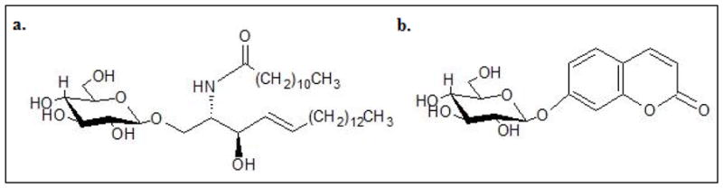
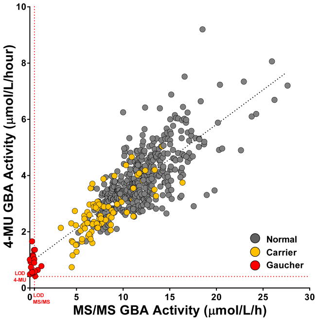
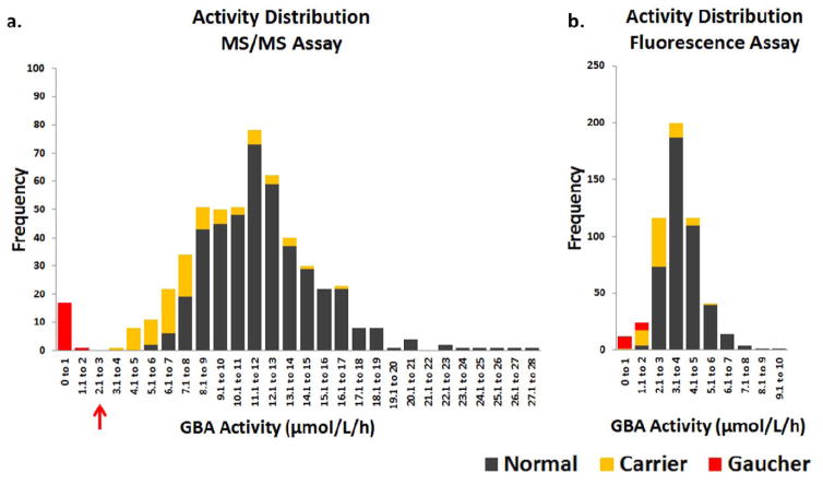
Deficiency of β-Glucocerebrosidase (GBA) activity causes Gaucher Disease (GD). GD can be diagnosed by measuring GBA activity (Beutler and Kuhl, 1990). In this study, we assayed dried blood spots from a cohort (n=528) enriched for GBA mutation carriers (n=78) and GD patients (n=18) using both the tandem mass spectrometry (MS/MS) and fluorescence assays and their respective synthetic substrates. The MS/MS assay differentiated normal controls, which included GBA mutation carriers, from GD patients with no overlap. The fluorescence assay did not always differentiate normal controls including GBA mutation carriers from GD patients and false positives were observed. The MS/MS assay improved specificity compared to the fluorescence assay.
Keywords: Dried blood spots (DBS); Gaucher disease (GD); Glucocerebroside; Lysosomal storage disorder (LSD); Newborn screening (NBS); β-Glucocerebrosidase (GBA).
Copyright © 2017 The Authors. Published by Elsevier Inc. All rights reserved.
Figures
References
-
- Beutler E, Kuhl B. In: Beta-glucosidase. 4. Williams WJ, et al., editors. McGraw-Hill Information Services Co., Health Professions Division; 1990.
-
- Grabowski GA, et al. Gaucher Disease: Phenotypic and Genetic Variation. In: Beaudet AL, et al., editors. The Online Metabolic and Molecular Bases of Inherited Disease. The McGraw-Hill Companies, Inc; New York, NY: 2014.
-
- Meikle PJ, et al. Prevalence of lysosomal storage disorders. Jama. 1999;281(3):249–54. - PubMed
-
- Weinreb NJ, et al. Prevalence of type 1 Gaucher disease in the United States. Arch Intern Med. 2008;168(3):326–7. author reply 327–8. - PubMed
Publication types
MeSH terms
Substances
Grants and funding
LinkOut - more resources
Full Text Sources
Other Literature Sources
Medical
Research Materials
Miscellaneous
