. 2017 Nov 3;2(17):eaam8834.
doi: 10.1126/sciimmunol.aam8834.

Oral epithelial cells orchestrate innate type 17 responses to Candida albicans through the virulence factor candidalysin

Affiliations

Oral epithelial cells orchestrate innate type 17 responses to Candida albicans through the virulence factor candidalysin

Akash H Verma et al. Sci Immunol. .

Abstract

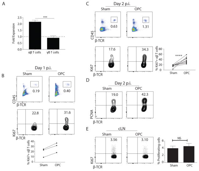
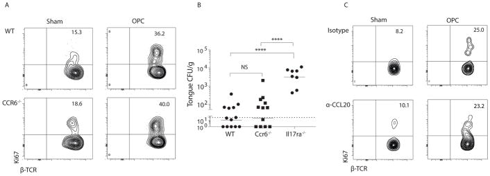
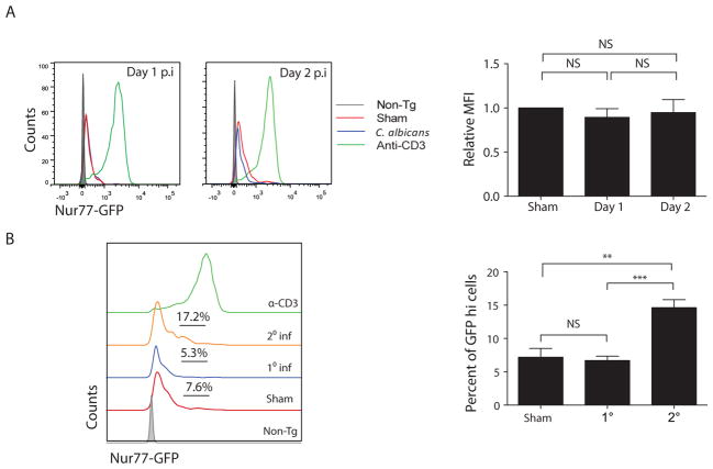
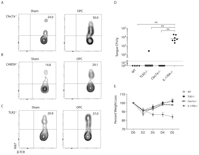
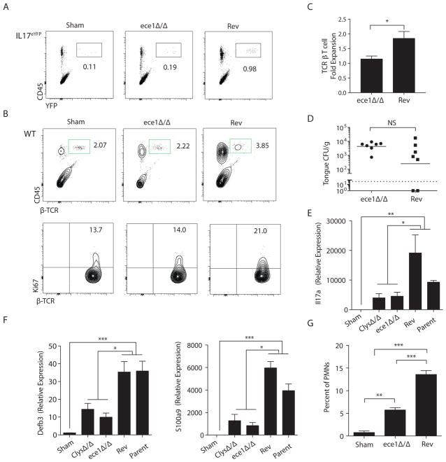
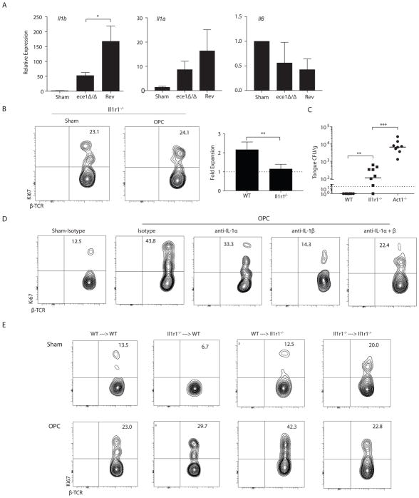
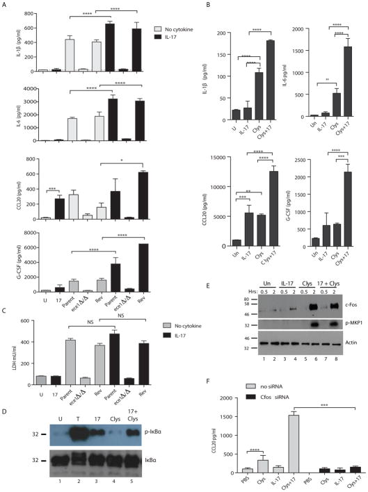
Candida albicans is a dimorphic commensal fungus that causes severe oral infections in immunodeficient patients. Invasion of C. albicans hyphae into oral epithelium is an essential virulence trait. Interleukin-17 (IL-17) signaling is required for both innate and adaptive immunity to C. albicans During the innate response, IL-17 is produced by γδ T cells and a poorly understood population of innate-acting CD4+ αβ T cell receptor (TCRαβ)+ cells, but only the TCRαβ+ cells expand during acute infection. Confirming the innate nature of these cells, the TCR was not detectably activated during the primary response, as evidenced by Nur77eGFP mice that report antigen-specific signaling through the TCR. Rather, the expansion of innate TCRαβ+ cells was driven by both intrinsic and extrinsic IL-1R signaling. Unexpectedly, there was no requirement for CCR6/CCL20-dependent recruitment or prototypical fungal pattern recognition receptors. However, C. albicans mutants that cannot switch from yeast to hyphae showed impaired TCRαβ+ cell proliferation and Il17a expression. This prompted us to assess the role of candidalysin, a hyphal-associated peptide that damages oral epithelial cells and triggers production of inflammatory cytokines including IL-1. Candidalysin-deficient strains failed to up-regulate Il17a or drive the proliferation of innate TCRαβ+ cells. Moreover, candidalysin signaled synergistically with IL-17, which further augmented the expression of IL-1α/β and other cytokines. Thus, IL-17 and C. albicans, via secreted candidalysin, amplify inflammation in a self-reinforcing feed-forward loop. These findings challenge the paradigm that hyphal formation per se is required for the oral innate response and demonstrate that establishment of IL-1- and IL-17-dependent innate immunity is induced by tissue-damaging hyphae.


Conflict of interest statement

Competing Interests: SLG has received grants from Novartis and Janssen. Candidalysin is the subject of a submitted patent to JRN, BH, DM, JPR: Naglik J, Moyes D, Tang S, Hube B, Wilson D, Hofs S, Richardson J (2013) Peptides and binding partners there for. WO 2014167335, Priority 2013-04-11.

Figures

Figure 1
Figure 1. Proliferation of oral TCRαβ+ cells following C. albicans infection
(A) Il17aeYFP mice (17) were challenged sublingually with PBS (sham) or C. albicans. Homogenates were prepared from pooled tongues (n=2–3). YFP+TCRαβ+ or YFP+TCRγδ+ cells in the CD45+CD4+ gate were assessed by flow cytometry. Data show fold-increase versus sham, pooled from 3–4 independent experiments. (B–C) WT mice (C57BL/6J) were infected with C. albicans, and tongue homogenates prepared on days 1 or 2 p.i. Cells were gated on lymphocytes and staining of CD45 and TCRβ is shown (top). Proliferation of CD45+CD4+TCRβ+ cells was determined by staining for Ki67 (bottom). Data representative of 10 experiments. Graph in C: mean ± SEM of proliferating TCRαβ+ cells on days 1 and 2. (D) WT mice were infected with C. albicans and tongue homogenates prepared on day 2 p.i. Proliferation was determined by PCNA staining. Data representative of 3 experiments. (E) WT cervical LNs were harvested on day 2 p.i. Proliferation of CD45+CD4+TCRβ+ cells was determined by anti-Ki67 staining. Graph shows mean ± SEM of Ki67+ CD4+ cells in cLNs. Data are representative of 2 experiments. Statistical analyses: Student’s t test or 1-way ANOVA.
Figure 2
Figure 2. CCR6 is dispensable for expansion of innate TCRαβ+ cells in oral candidiasis
(A) WT or Ccr6−/− mice were infected with C. albicans, and proliferation of oral TCRαβ+ cells was determined at day 2 p.i. Data representative of 3 independent experiments. (B) Indicated mice were infected orally with C. albicans, and fungal burden was assessed by CFU enumeration on day 4 p.i.. Bars = geometric mean. Each point represents an individual mouse. Dashed line = limit of detection (LOD, 30 CFU) (52). Data are compiled from 4 independent experiments. (C) WT mice were injected with 100 ug anti-CCL20 Abs or isotype controls on day -1 relative to infection. Proliferation of TCRαβ+ cells was determined on day 2 p.i. Data are representative of 2 experiments. Student’s t test or ANOVA used for statistics.
Figure 3
Figure 3. A primary C. albicans infection activates innate TCRαβ+ cells without engaging the TCR
(A) Nur77GFP mice were sham-treated (red line) or infected with C. albicans and tongue homogenates prepared on days 1 or 2 p.i (blue line). Controls received anti-CD3 Abs (green line) to stimulate the TCR on all T cells. WT (“non-Tg”) mice were negative controls for GFP staining (grey line). Left: fluorescence intensity of GFP in oral CD45+CD4+TCRβ+ cells. Right: Relative mean fluorescence intensity (MFI) of GFP in CD45+CD4+TCRβ+ cells was assessed and normalized to sham. NS, not significant. Data from 3 independent experiments. (B) Nur77GFP mice were infected with C. albicans. Tongue homogenates were prepared 2 d p.i. (“1° Inf”). To induce C. albicans-specific TCR signaling (“2° Inf”), mice were infected orally, rested for 6 weeks, then re-challenged with a second oral infection (8). Left: GFP fluorescence in oral CD45+CD4+TCRβ+ cells, with % GFPhi cells indicated. Green line shows staining in mice administered agonistic anti-CD3 Abs, as in panel A. Right: Compiled percentage of GFPhi cells per cohort. Data representative of 3–4 independent experiments. Graphs show mean + SEM, analyzed by student’s t test or ANOVA.
Figure 4
Figure 4. TLR2 and Dectin-1 are dispensable for C. albicans-induced proliferation of innate TCRαβ+ cells
(A–C) Indicated mice were infected with C. albicans and proliferation of CD45+CD4+TCRβ+ cells determined on day 2 p.i. Data representative of 2–3 independent experiments. (D) Indicated mice were infected and fungal burden quantified on day 5 p.i. Bars = geometric mean. Dashed line = LOD. Data from 2 independent experiments. Data analyzed by ANOVA, Mann Whitney correction. (E) Average % weight loss is shown.
Figure 5
Figure 5. Candidalysin drives proliferation of innate IL-17-producing TCRαβ+ cells
(A) Il17aeYFP mice were infected with C. albicans (ece1Δ/Δ or Revertant “Rev”) and homogenates prepared 2 d p.i. Staining of CD45 and YFP in lymphocyte gate is shown. Data representative of 2 experiments. (B) WT mice were infected with the indicated strains of C. albicans, and expansion (top) and proliferation (bottom) of oral TCRαβ+ cells was analyzed at day 2 p.i.. Data representative of 3 experiments. (C) Fold-expansion of TCRβ+ cells following infection with ece1Δ/Δ or Rev strains. Data pooled from 4 experiments. (D) Fungal loads were assessed at day 2 p.i.. Bar = geometric mean. Data pooled from 2 experiments. (E–F) Tongue homogenates were prepared 2 days after infection with the indicated C. albicans strains. Total mRNA was subjected to qPCR normalized to Gapdh. Graphs show mean + SEM normalized to sham. Data compiled from 7–8 mice per group from 2 independent experiments. (G) Percentage of CD11b+Ly6Ghi cells in tongue was assessed at day 2 p.i. + SEM, compiled from 3 experiments. Statistics analyzed by student’s t test or ANOVA.
Figure 6
Figure 6. IL-1 activates innate TCRαβ+ T cell proliferation and anti-fungal immunity in a T cell intrinsic and T cell extrinsic manner
(A) WT mice were infected with the indicated C. albicans strains and gene expression was measured on day 2 p.i.. Data show mean + SEM normalized to sham, from 7–8 mice/group in 2 experiments. (B) Expansion and proliferation of TCRαβ+ cells in Il1r1−/− mice at day 2 p.i. Data are from 3 experiments. (C) Fungal burdens in the indicated mice were quantified on day 5 p.i., from 2 experiments. (D) WT mice were administered anti-IL-1α, anti-IL-1β or isotype control Abs (1.0 mg/mouse used alone or 0.5 mg each when used together) on day -1 p.i.. Proliferation of oral TCRαβ+ cells was assessed at day 2 day p.i. Data representative of 2 experiments. (E) Reciprocal adoptive transfers of femoral BM were performed in WT or Il1r1−/− mice and proliferation of oral TCRαβ+ cells was determined. Experimental chimera results are representative of 2 experiments; control chimera data are from 1 experiment. Data analyzed by Student’s t test or ANOVA.
Figure 7
Figure 7. Candidalysin and IL-17 signal synergistically or additively in oral epithelial cells
(A) TR146 OECs were untreated (“U”, grey bars) or stimulated with IL-17 (200 ng/ml, black bars). Cells were infected with WT C. albicans (Bwp17+CIp30, “Parent”), ece1Δ/Δ or the Revertant (Rev) for 24 h. Supernatants were analyzed by Luminex (IL-1β, IL-6, G-CSF) or ELISA (CCL20). Graphs indicate mean + SEM. Data are representative of 2 experiments. (B) TR146 cells were untreated (“U”, grey bars) or treated with IL-17 (200 ng/ml, black bars) or Candidalysin peptide (Clys, 15 μM) for 24 h and analyzed as in panel A. (C) TR146 cells were incubated with C. albicans ± IL-17 (200 ng/ml). LDH in supernatants was evaluated after 24 h, representative of 3 experiments. (D) TR146 cells were treated with TNFα (20 ng/ml), IL-17 (200 ng/ml) or Candidalysin (15 μM) for 5 min. Lysates were immunoblotted for phospho-IκBα and total IκBα. (E) TR146 cells were incubated with TNFα (20 ng/ml), IL-17 (200 ng/ml) or Candidalysin (15 μM) for 30 min or 2 h. Lysates were immunoblotted for c-Fos, phospho-MKP1 or Actin. Data are representative of 2 experiments. (F) TR146 cells were transfected with c-Fos siRNA and stimulated for 24 h with PBS, Clys or IL-17. Supernatants were assessed for CCL20 by ELISA. Data are representative of 2 independent experiments. All data analyzed by ANOVA and Student’s t-test.

Comment in

  • Candidalysin sets off the innate alarm.
    Li X, Leonardi I, Iliev ID. Li X, et al. Sci Immunol. 2017 Nov 3;2(17):eaao5703. doi: 10.1126/sciimmunol.aao5703. Sci Immunol. 2017. PMID: 29101210 Free PMC article.

Similar articles

Cited by

References

    1. Brown GD, et al. Hidden killers: human fungal infections. Sci Transl Med. 2012;4:165rv113. - PubMed
    1. Jabra-Rizk MA, et al. Candida albicans Pathogenesis: Fitting Within the “Host-Microbe Damage Response Framework”. Infect Immun. 2016;84:2724–2739. - PMC - PubMed
    1. Conti HR, Gaffen SL. IL-17-Mediated Immunity to the Opportunistic Fungal Pathogen Candida albicans. J Immunol. 2015;195:780–788. - PMC - PubMed
    1. Milner J, Holland S. The cup runneth over: lessons from the ever-expanding pool of primary immunodeficiency diseases. Nat Rev Immunol. 2013;13:635–648. - PubMed
    1. Conti H, et al. Th17 cells and IL-17 receptor signaling are essential for mucosal host defense against oral candidiasis. J Exp Med. 2009;206:299–311. - PMC - PubMed

MeSH terms