. 2017 Nov 3;7(1):14487.
doi: 10.1038/s41598-017-13751-z.

In vitro selections of mammaglobin A and mammaglobin B aptamers for the recognition of circulating breast tumor cells

Affiliations

In vitro selections of mammaglobin A and mammaglobin B aptamers for the recognition of circulating breast tumor cells

Eman M Hassan et al. Sci Rep. .

Abstract

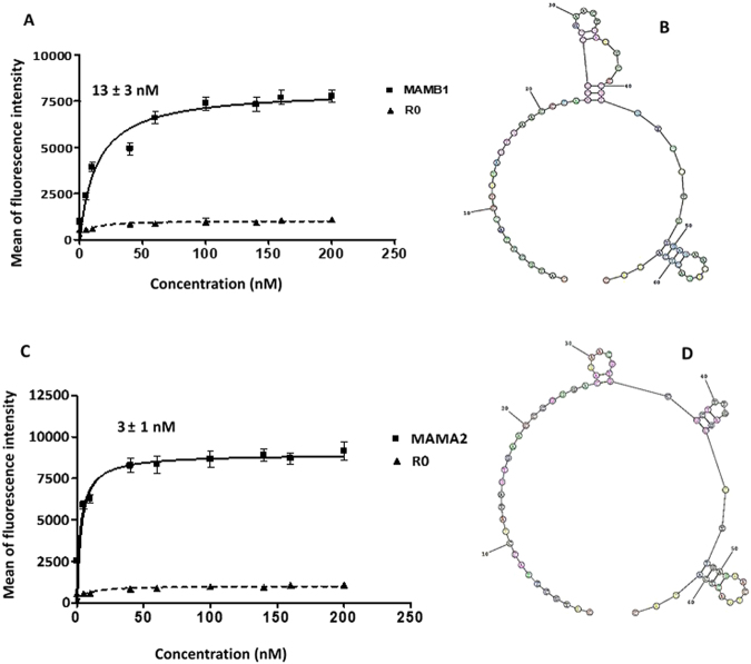
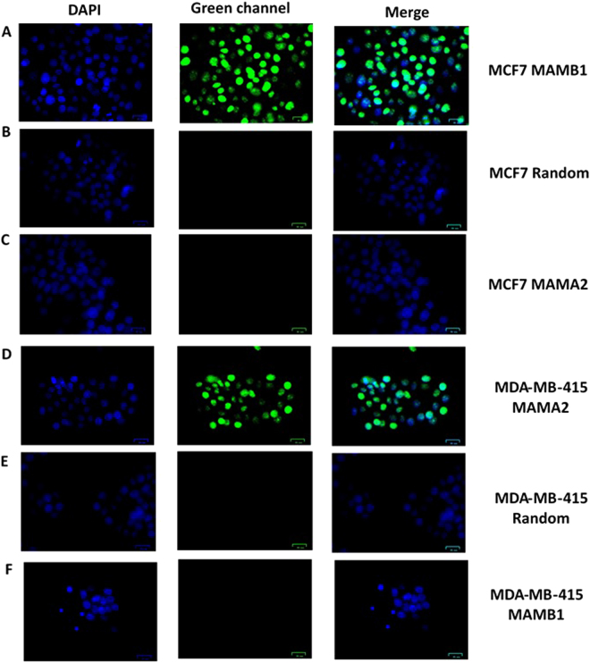
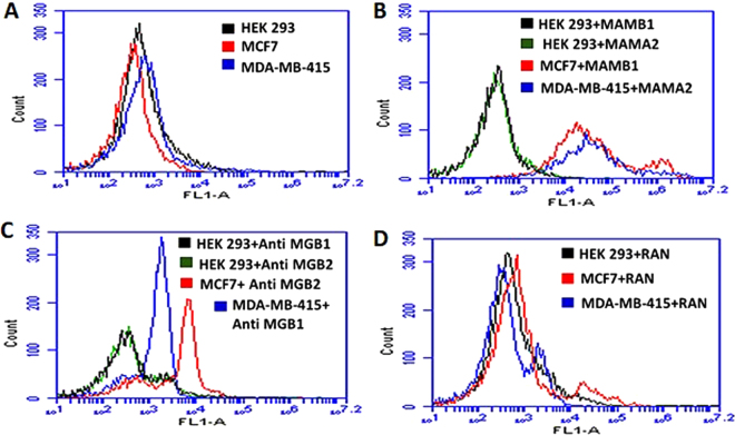
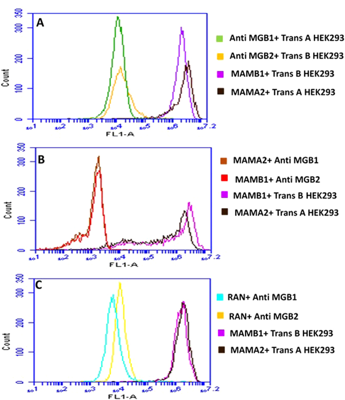
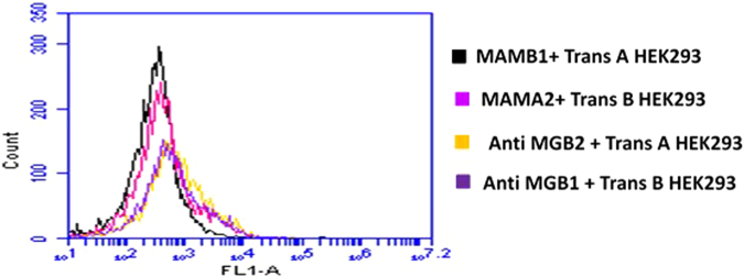
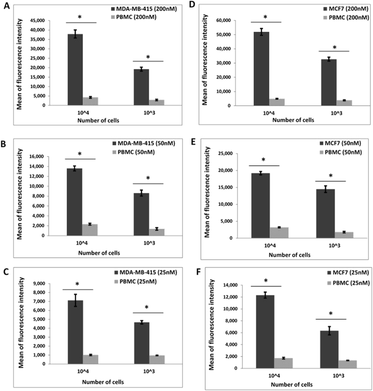
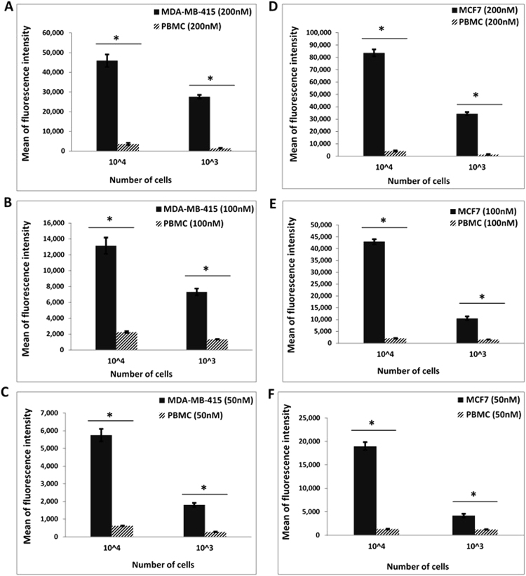
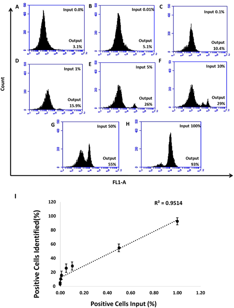
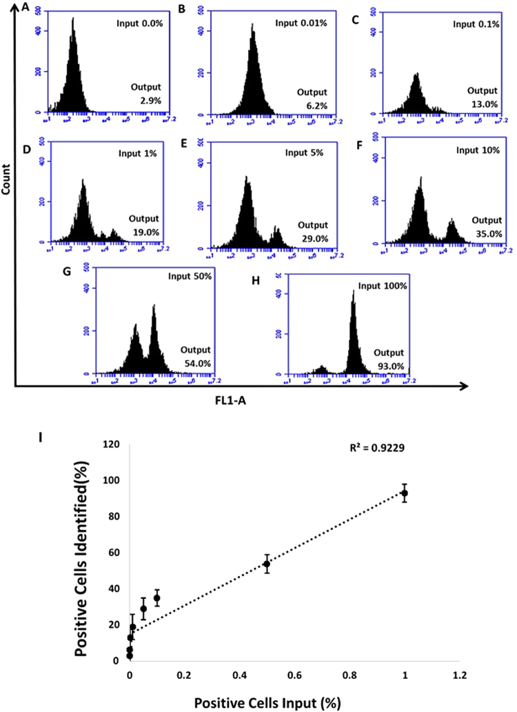
Mammaglobin B (MGB2) and mammaglobin A (MGB1) are proteins expressed in metastatic breast cancers. The early detection of circulating tumor cells (CTCs) in breast cancer patients is crucial to decrease mortality rate. Herein, novel aptamers were successfully selected and characterized against MGB2 and MGB1 proteins using a hybrid SELEX approach. The potential use of the selected aptamers in breast CTC detection was studied using spiked breast cancer cells in whole blood lysate. The results obtained from this study showed that the selected aptamers (MAMB1 and MAMA2) bind to their target breast cancer cell lines with high affinity (low nanomolar Kd values) and specificity. They also bind to their free recombinant target proteins and show minimal non-specific binding to normal and other cancer cell lines. Additionally, they were able to distinguish a low number of breast cancer cells spiked in whole blood lysate containing normal blood cells. The results obtained in this study indicate the great potential for the use of aptamers to detect MGB1 and MGB2 protein biomarkers, expressed on the surface of breast CTCs.


Conflict of interest statement

The authors declare that they have no competing interests.

Figures

Figure 1
Figure 1
Schematic diagram of hybrid SELEX method for selection of MGB2 and MGB1-specific DNA aptamers. Hybrid SELEX is divided into (A) a protein-based SELEX in which GST-MGB2 conjugated beads (brown) and GST-MGB1 proteins conjugated beads (purple) were used. The protein SELEX was done for 21 rounds for MGB2 and 4 round for MGB1. Subsequently, the enriched pools of both proteins were transferred to cells (B) to start cell-based SELEX in which HCAEC and MCF10A were used as counter selection cell lines. MCF7 was used as the target selection cell line for MGB2 and MDA-MB-415 was used as the target selection cell line for MGB1. After 7 rounds of cell-SELEX to each target cell line, the final pools for both targets were transferred back to the proteins for another 2 additional rounds for each MGB2 and MGB1 to make the library more specific to MGB2 and MGB1.
Figure 2
Figure 2
Monitoring the enrichment of ssDNA library of MGB2 and MGB1 during hybrid SELEX. (A) Percentage recovery of MGB2 library during protein SELEX. GST-protein (blue) was used for counter selection throughout the selection. MGB1 protein (green) was used as the counter selection target in R16 to R18 to split the main library into two parts to eliminate the sequences that could bind to MGB1 and MGB2 targets together. (B) Percentage recovery of MGB1 protein library during protein SELEX. R17 counter selection (green) from MGB2 SELEX was used as the initial ssDNA library of MGB1 SELEX. The percentage recovery for both proteins SELEX was measured as the emission of FAM labelled ssDNA libraries at 520 nm. (C) The fluorescence intensity (FL1-A) of MCF7 (MGB2 positive cell line) when binding to R0, R21, and R30 of MGB2. (D) The fluorescence intensity (FL1-A) of MDA-MB-415 (MGB1 positive cell line) when binding to R0, R4, and R13 of MGB1. (E,F) The fluorescence intensity (FL1-A) of HCAEC cells and MCF10A (counter cell lines) respectively when binding to R0, R21 MGB2, R4 MGB1, R30 MCF7 and R13 MDA-MB-415. The fluorescence intensity was measured using flow cytometry by incubating the 6-FAM tagged ssDNA with the cells for 30 min at 4 °C in PBS. The fluorescence intensity of cells from all cell lines (control/background signal) was subtracted from the fluorescence intensity of the positive and counter cell lines and included in the graph.
Figure 3
Figure 3
Screening results of the aptamer candidates against cell-SELEX cell lines using flow cytometry. (A) Screening results of MGB2 aptamers show six aptamers out of twelve (MAMB0, MAMB1, MAMB4, MAMB8, MAMB12, MAMB57) with higher fluorescence intensity in the target selection cell line than counter selection ones. (B) Screening results of MGB1 aptamers show four aptamers out of eight (MAMA2, MAMA5, MAMA6, and MAMA12) with higher fluorescence intensity in the target selection cell line than counter selection ones. Promising aptamers were chosen for further studies. The experiments were repeated twice. Values are shown as means ± S.E.M.
Figure 4
Figure 4
Binding curves of 6-FAM-labeled aptamers to MCF7 and MDA-MB-415 target cell lines. (A) MAMB1 Kd curve: MCF7 cells were incubated with increasing concentrations (nM) of MAMB1 then was evaluated by flow cytometry. (B) The predicted secondary structure of MAMB1 aptamer using RNAstructure software. (C) MAMA2 Kd curve: MDA-MB-415 cells were incubated with increasing concentrations (nM) of MAMA2 then was evaluated by flow cytometry. (D) The predicted secondary structure of MAMA2 aptamer using RNAstructure software. To calculate the apparent Kd of the interaction of MAMB1 and MAMA2 to their corresponding cell lines, the mean fluorescence intensity of the aptamer-cell dissociation vs. concentration was fit to the equation Y=Bmax X/(Kd+X). R0 ssDNA library was used as control. The Kd graphs are the average of three trials. Values are shown as means ± S.E.M.
Figure 5
Figure 5
Fluorescence microscopy images of aptamers MAMB1 and MAMA2 binding to target breast cancer cell lines. (A) MAMB1 binding to MCF7 cells, (B) random sequence binding to MCF7, (C) MAMA2 binding to MCF7. (D) MAMA2 binding to MDA-MB-415 cells, (E) random sequence binding to MDA-MB-415, (F) MAMB1 binding to MDA-MB-415. Both aptamers and the random sequence were incubated with the cells for 30 min and then imaged. Scale bars correspond to 25 μm in all images.
Figure 6
Figure 6
Binding of MAMB1, MAMA2 aptamers and MGB2, MGB1 antibodies to nontransfected HEK293, MCF7, and MDA-MB-415 cells. (A) Mean of fluorescence intensity of non-transfected HEK293 cells (black), MCF7 (red), and MDA-MB-415 (blue) without probes (cells only). (B) Mean of fluorescence intensity of non-transfected HEK293 cells binding to MAMB1 (black), MAMA2 (green), MCF7 binding to MAMB1 (red), and MDA-MB-415 binding to MAMA2 (blue). (C) Mean of fluorescence intensity of non-transfected HEK293 cells binding to anti-MGB1 antibody (black), anti MGB2 antibody (green), MCF7 binding to anti MGB2 antibody (red), and MDA-MB-415 binding to MGB1 antibody (blue). (D) Mean of fluorescence intensity of non-transfected HEK293 cells (black), MCF7 (red), and MDA-MB- 415 (blue) binding to a random sequence. Cells were incubated with their corresponding probes and 10000 events was counted by flow cytometry. Then data was analyzed using flow cytometry C6 sampler software.
Figure 7
Figure 7
Competition binding assays of MAMB1, MAMA2 aptamers and anti MGB, anti MGB1 antibodies on transfected HEK293 cells with MGB2 and MGB1 plasmids. (A) Mean of fluorescence intensity of transfected HEK293 with MGB2 plasmid (Trans B) binding to anti MGB2 (orange) and MAMB1 (purple), and transfected HEK293 with MGB1 plasmid (Trans A) binding to anti MGB1 (green) and MAMA2 (brown). (B) Mean of fluorescence intensity of Trans B and Trans A HEK293 blocked with unlabeled MAMB1 and MAMA2 aptamers respectively, and probed with anti MGB2 (red) in the case of Trans B, and anti MGB1 (brown) in the case of Trans A. Mean of fluorescence intensity of Trans B and Trans A binding to MAMB1 (purple), and MAMA2 (brown) same as in A. (C) Mean of fluorescence intensity of Trans B and Trans A HEK293 blocked with random sequence (RAN) and probed with anti MGB2 (dark yellow) in the case of Trans B and anti MGB1 (light blue) in the case of Trans A. Mean of fluorescence intensity of Trans B and Trans A binding to MAMB1 (purple) and MAMA2 (brown) same as in A. Cells were incubated with their corresponding probes and 10000 events was counted by flow cytometry. Then data was analyzed using flow cytometry C6 sampler software.
Figure 8
Figure 8
Selectivity test performed on transfected HEK293 cells with MGB2 and MGB1 plasmids. The figure shows the mean of fluorescence intensity of MAMB1 (black), MAMA2 (pink), anti MGB2 (dark yellow), and anti MGB1 (purple) binding to Trans A and Trans B HEK293 cells respectively. Cells were incubated with their counter probes transfected HEK293, and 10000 events was counted by flow cytometry. Then data was analyzed using flow cytometry C6 sampler software.
Figure 9
Figure 9
Binding of MAMB1 and MAMA2 aptamers to their breast cancer cells in plasma by flow cytometry. (A-C) Mean of fluorescence intensity of MDA-MB-415 cancer cells and PBMC normal cells at 200nM (A), 50nM (B), and 25nM (C) concentrations of MAMA2 aptamer. (D–F) Mean of fluorescence intensity of MCF7 cancer cells and PBMC normal cells at 200nM (D), 50nM (E), and 25nM (F) concentrations of MAMB1 aptamer. Two cell numbers from both cancer and normal cells were used: 104 and 103 cells. Values are shown as means ± S.E.M. of three trials. The statistical significance was determined by one way ANOVA followed by Fisher LSD multiple tests using SPSS software (SPSS, version 23). P Values less than 0.05 were considered to be significantly different.
Figure 10
Figure 10
Binding of MAMB1 and MAMA2 aptamers to their breast cancer cells in whole Blood lysate by flow cytometry. (AC) Mean of fluorescence intensity of MDA-MB-415 cancer cells and PBMC normal cells at 200 nM (A), 100 nM (B) and 50 nM (C) concentrations of MAMA2 aptamer. (D–F) Mean of fluorescence intensity of MCF7 cancer cells and PBMC normal cells at 200nM (D), 100nM (E) and 50nM (F) concentrations of MAMB1 aptamer. Two cell numbers from both cancer and normal cells were used: 104 and 103 cells. Values are shown as means ± S.E.M. of three trials. The statistical significance was determined by one way ANOVA followed by Fisher LSD multiple tests using SPSS software (SPSS, version 23). P Values less than 0.05 were considered to be significantly different.
Figure 11
Figure 11
Selective recognition of MCF7 cells in mixed cell samples with the 6-FAM labelled MAMB1 aptamer in whole blood lysate. Cell mixture samples containing MCF7 and PBMC were prepared in different percentages of MCF7 ((A–G) 0%, 0.01%, 0.1%, 1%, 5%, 10%, 50%, and 100%). (I) plot of the percentage of cancer cells (MCF7) spiked (X-axis) and the percentage of positive cells identified (Y-axis). Values are shown as means ± S.E.M. of three trials. The statistical significance was determined by one way ANOVA followed by Fisher LSD multiple tests using SPSS software (SPSS, version 23). P Values less than 0.05 were considered to be significantly different.
Figure 12
Figure 12
Selective recognition of MDA-MB-415 cells in mixed cell samples with the 6-FAM labelled MAMA2 aptamer in whole blood lysate. Cell mixture samples containing MDA-MB-415 and PBMC were prepared in different percentages of MDA-MB-415 ((AG) 0%, 0.01%, 0.1%, 1%, 5%, 10%, 50%, and 100%). (I) Plot of the percentage of cancer cells (MDA-MB-415) spiked (X-axis) and the percentage of positive cells identified by the MAMA2 aptamer (Y-axis). Values are shown as means ± S.E.M. of three trials. The statistical significance was determined by one way ANOVA followed by Fisher LSD multiple tests using SPSS software (SPSS, version 23). P Values less than 0.05 were considered to be significantly different.

Similar articles

Cited by

References

    1. Hanahan D, Weinberg RA. Hallmarks of cancer: the next generation. Cell. 2001;144:646–674. doi: 10.1016/j.cell.2011.02.013. - DOI - PubMed
    1. Ferlay, J. et al. GLOBOCAN 2012 v1.0, Cancer Incidence and Mortality Worldwide: IARC CancerBase No. 11 [Internet]. International Agency for Research on Cancer. Available on: http://globocan.iarc.fr (2013).
    1. U.S. Breast cancer statistics. http://www.breastcancer.org/ (2016). - PubMed
    1. Redig AJ, McAllister SS. Breast cancer as a systemic disease: a view of metastasis. J. Intern. Med. 2013;274(2):113–126. doi: 10.1111/joim.12084. - DOI - PMC - PubMed
    1. Vanio, H., Bianchini, F. IARC Handbooks of Cancer Prevention, Volume 7: Breast Cancer Screening. 4–16 (IARC Press, France, 2002).

Publication types