. 2017 Nov 3;7(1):14500.
doi: 10.1038/s41598-017-14202-5.

T cells upon activation promote endothelin 1 production in monocytes via IFN-γ and TNF-α

Affiliations

T cells upon activation promote endothelin 1 production in monocytes via IFN-γ and TNF-α

Shoshi Shinagawa et al. Sci Rep. .

Abstract

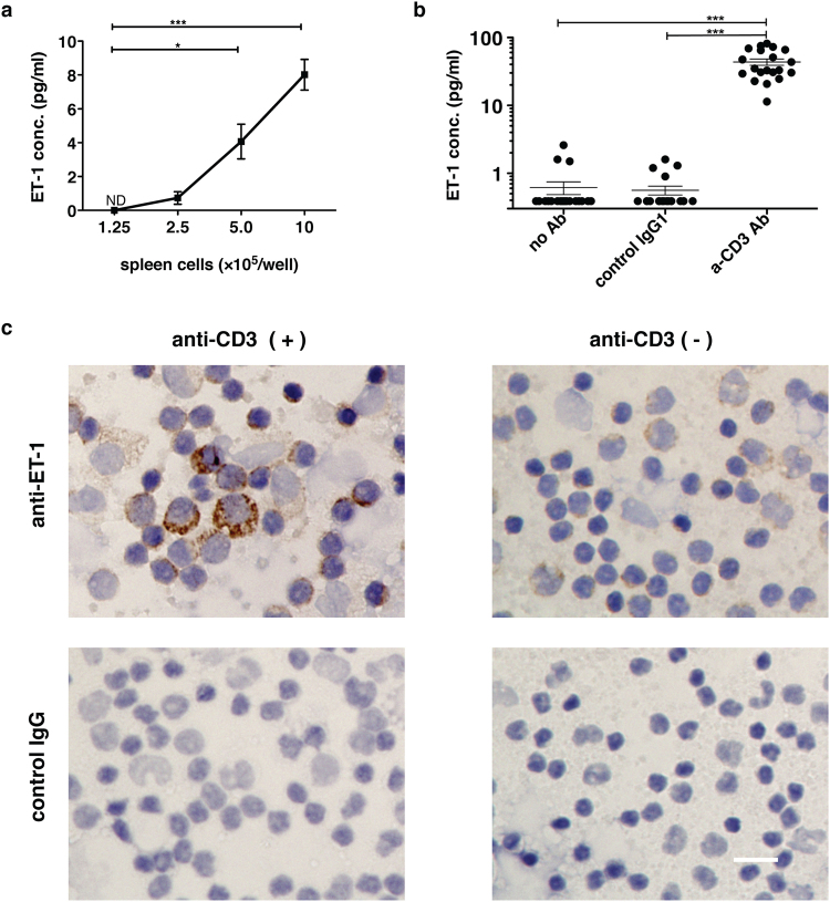
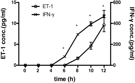
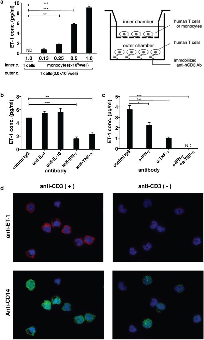
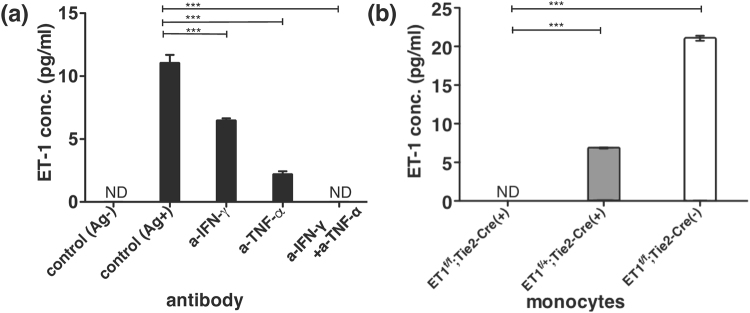
Endothelin 1 (ET-1), mainly produced from vascular endothelial cells, induces vasoconstriction in physiological conditions. The endothelin receptor antagonist is among the most effective agents for pulmonary hypertension. However, little is known about the production source of ET-1 in inflammation and immunity. Here, we studied whether T cell-mediated ET-1 production system exists and operates independent of the production system in vascular endothelial cells. ET-1 production was readily detectable in the culture supernatant of human PBMCs and murine spleen cells stimulated with anti-CD3 antibody. Immunocytostaining showed that ET-1-producing cells emerged only in PBMCs stimulated with anti-CD3 antibody. Using the Transwell system, both murine and human monocytes sorted with magnetic beads in the inner chamber produced ET-1 when T cells were activated with antigen or anti-CD3 antibody in the outer chamber. This ET-1 production was inhibited by anti-IFN-γ and/or TNF-α antibody. Furthermore, monocytes purified from ETflox/flox;Tie2-Cre( + ) mice, which conditionally lack ET-1 in hematopoietic stem cells and vascular endothelial cells, did not produce ET-1 even when stimulated by antigen-specific T cell activation. This study demonstrates the existence of an immune-mediated ET-1 production induced by T cells upon activation through IFN-γ and TNF-α.


Conflict of interest statement

The authors declare that they have no competing interests.

Figures

Figure 1
Figure 1
ET-1 production by immune cells following anti-CD3 Ab-mediated activation of T cells in murine spleen cells and human PBMCs. ET-1 production in the culture supernatant of 1 × 106 cells/well of (a) murine spleen cells (b) human PBMCs from 20 healthy donors cultured for 24 h with or without stimulation with 10 μg/ml immobilized anti-human CD3 Ab or control IgG1. The value under the limit of detection (0.39 pg/ml) with ET-1 ELISA assay was defined as 0.39 pg/ml. The assays were performed in triplicate wells. Data are expressed as the mean ± SEM. *p < 0.05, ***p < 0.001 as compared with negative control by (a) one-way ANOVA followed by post-hoc Tukey’s multiple comparison test and (b) Kruskal-Wallis followed by post-hoc Dunn’s multiple comparison test. (c) Representative images of immunocytochemical staining of human PBMCs with anti-human ET1 mAb. PBMCs were cultured in the presence or absence of immobilized anti-human CD3 Ab for 28 h. Brefeldin A (10 μg/ml) was added to the culture for the last 4 h. After fixation with 4% paraformaldehyde and methanol, DAB staining was performed using anti-hET1 Ab (TR.ET.48.5) or control IgG. Scale bar represents 20 μm. Similar results (a) and images (c) were obtained in 3 different experiments.
Figure 2
Figure 2
Comparison of the production kinetics between IFN-γ and ET-1 in the culture supernatant of PBMCs stimulated with immobilized anti-CD3 Ab. ET1 production in the culture supernatant of 1 × 106 cells/well of human PBMCs from a healthy donor cultured with 10 μg/ml immobilized anti-human CD3 Ab. The culture supernatants at appropriate times were collected and measured by ET-1 or IFN-γ ELISA. Assays were performed in triplicate wells, and data represent means ± SEM. ND = not detected. * P < 0.001 as calculated compared with negative control (Time 0) by two-way ANOVA followed by post-hoc Sediak’s multiple comparison test. Similar results were obtained in 3 different experiments.
Figure 3
Figure 3
The relationship between T cells and monocytes in ET-1 induction with the double chamber system using human cells. After PBMCs were prepared from 100 ml of whole blood of a volunteer, T cells and monocytes were purified from human PBMCs with Mini-MACS, respectively. Three million T cells were stimulated with immobilized anti-CD3 Ab in the outer chamber of the Transwell system (See the schema on the right of Fig. 3a.). In the inner chamber (a), the number of purified T cells or monocytes with magnetic beads is shown on the x axis. For panels (b) and (c), 1.5 × 106 purified human monocytes were cultured for 24 h. The ET-1 concentration in the supernatant of each culture was measured using ELISA. The concentration of each blocking Ab used was 30 µg/ml. ND = not detected. The assays were performed in triplicate wells. Data are expressed as the mean ± SEM. *p < 0.05, **p < 0.01, ***p < 0.001 as compared with negative control with T cells in the inner chamber (a) or positive control (b,c) by one-way ANOVA followed by post-hoc Tukey’s multiple comparison test. (d) Images of the detection of ET-1 production from monocytes purified from PBMCs in the inner chamber. The monocytes in the inner chamber were cultured with T cells in the outer chamber in the presence or absence of immobilized anti-human CD3 Ab for 28 h. Brefeldin A (10 μg/ml) was added to the culture for the last 4 h. After fixation with 4% paraformaldehyde and 0.5% Triton X-100, immunofluorescent staining was performed using anti-ET-1 Ab (TR.ET.48.5) with anti-polyclonal mouse IgG(H + L) conjugated with Alexa Fluor 594 (Red) and anti-human CD14 (1:25 dilution) conjugated with Alexa Fluor 488 (Green). Nuclei were visualized with DAPI (Blue). Similar results and images were obtained in at least 3 different experiments (ad).
Figure 4
Figure 4
Effect of antigen-specific activation of T cells on monocytes in mice. Three million Tax11-19-specific CTLs were stimulated with 4 × 106 mitomycin C-treated syngeneic spleen cells from HHD mice in the presence or absence of 0.1 μM Tax11-19 peptide as an antigen (Ag) in the outer chamber. In the inner chamber, after bone marrow cells were prepared from 4 mice of femurs in each strain, 1.5 × 106 /well of monocytes purified with Mini-MACS from bone marrow cells of (a) HHD mice and (b) ET1flox/flox;Tie2-Cre(+), ET1flox/+;Tie2-Cre(+), and ET1flox/flox;Tie2-Cre(−) mice were cultured for 24 h. The concentration of each blocking Ab used was 30 µg/ml. ND = not detected. The assays were performed in triplicate wells. Data are expressed as the mean ± SEM. ***p < 0.001 as compared with positive (a) or negative (b) control by one-way ANOVA followed by post-hoc Tukey’s multiple comparison test. Similar results were obtained in two experiments (a,b).

References

    1. Davenport AP, et al. Endothelin. Pharmacological reviews. 2016;68:357–418. doi: 10.1124/pr.115.011833. - DOI - PMC - PubMed
    1. Yanagisawa M, et al. A novel potent vasoconstrictor peptide produced by vascular endothelial cells. Nature. 1988;332:411–415. doi: 10.1038/332411a0. - DOI - PubMed
    1. Kedzierski RM, Yanagisawa M. Endothelin system: the double-edged sword in health and disease. Annu Rev Pharmacol Toxicol. 2001;41:851–876. doi: 10.1146/annurev.pharmtox.41.1.851. - DOI - PubMed
    1. Maguire JJ, Davenport AP. Endothelin receptors and their antagonists. Seminars in nephrology. 2015;35:125–136. doi: 10.1016/j.semnephrol.2015.02.002. - DOI - PMC - PubMed
    1. Sampaio AL, Rae GA, Henriques M. Effects of endothelin ETA receptor antagonism on granulocyte and lymphocyte accumulation in LPS-induced inflammation. J Leukoc Biol. 2004;76:210–216. doi: 10.1189/jlb.1003504. - DOI - PubMed

Publication types

MeSH terms