. 2017 Nov 3;8(1):1305.
doi: 10.1038/s41467-017-01431-5.

piRNAs and Aubergine cooperate with Wispy poly(A) polymerase to stabilize mRNAs in the germ plasm

Affiliations

piRNAs and Aubergine cooperate with Wispy poly(A) polymerase to stabilize mRNAs in the germ plasm

Jérémy Dufourt et al. Nat Commun. .

Abstract

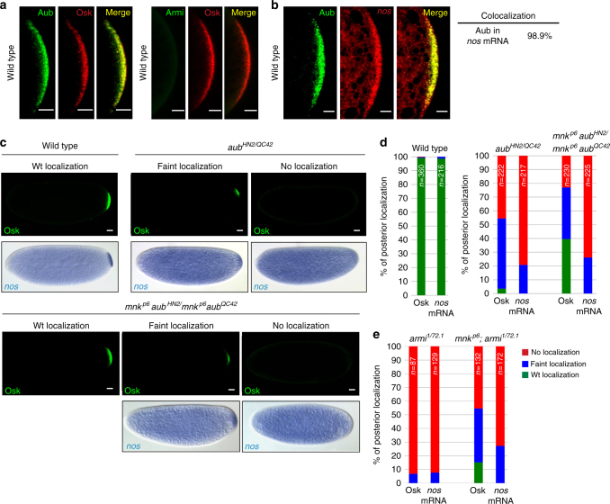
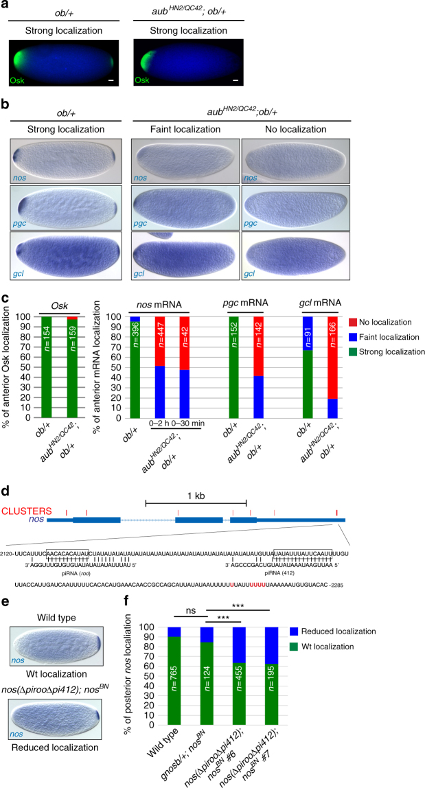
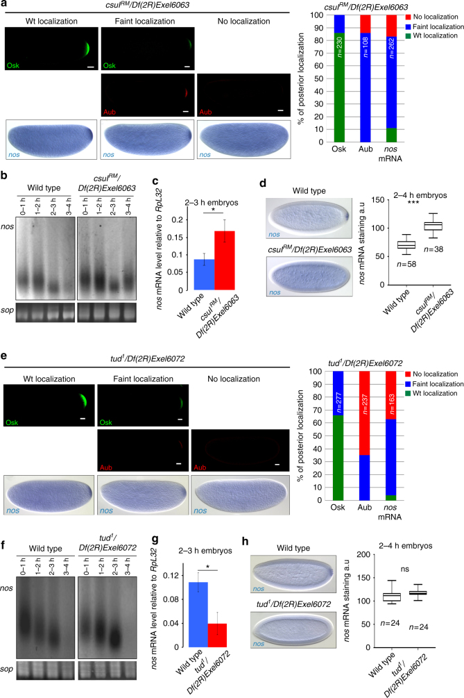
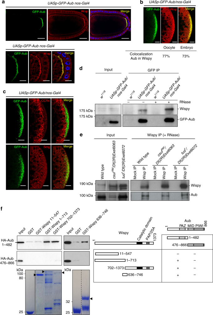
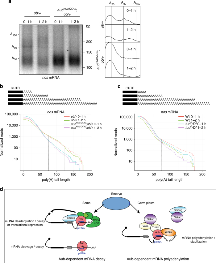
Piwi-interacting RNAs (piRNAs) and PIWI proteins play a crucial role in germ cells by repressing transposable elements and regulating gene expression. In Drosophila, maternal piRNAs are loaded into the embryo mostly bound to the PIWI protein Aubergine (Aub). Aub targets maternal mRNAs through incomplete base-pairing with piRNAs and can induce their destabilization in the somatic part of the embryo. Paradoxically, these Aub-dependent unstable mRNAs encode germ cell determinants that are selectively stabilized in the germ plasm. Here we show that piRNAs and Aub actively protect germ cell mRNAs in the germ plasm. Aub directly interacts with the germline-specific poly(A) polymerase Wispy, thus leading to mRNA polyadenylation and stabilization in the germ plasm. These results reveal a role for piRNAs in mRNA stabilization and identify Aub as an interactor of Wispy for mRNA polyadenylation. They further highlight the role of Aub and piRNAs in embryonic patterning through two opposite functions.


Conflict of interest statement

The authors declare no competing financial interests.

Figures

Fig. 1
Fig. 1
Aub and Armi have a direct role in nos mRNA posterior localization. a Immunostaining of wild-type 0–2 h-embryos with anti-Osk and either anti-Aub or anti-Armi. Posterior poles are shown. b Immuno-FISH of wild-type 0–2 h-embryos with anti-Aub and nos RNA probe. Quantification of colocalization using the Manders coefficient is indicated. Scale bars: 10 μm in a, b. c Immunostaining with anti-Osk and in situ hybridization with nos RNA probe of 0–2 h- wild-type, aub mutant or mnk aub double-mutant embryos. Scale bars: 30 μm. d, e Quantification of posterior localization of Osk and nos mRNA shown in c for aub and mnk aub mutant embryos, and in (Supplementary Fig. 1b) for armi and mnk; armi mutant embryos; the legend is in e. For all figures, the number of embryos (n) is indicated; the posterior pole is to the right
Fig. 2
Fig. 2
Role of Aub and piRNAs in germ cell mRNA localization to the germ plasm. a Immunostaining with anti-Osk of osk-bcd3′UTR (ob) embryos in wild-type or aub mutant backgrounds. The DAPI staining background (blue) shows the bulk of the embryo. Scale bars: 30 μm. b In situ hybrydization of 0–2 h ob/ + embryos either in wild-type or aub mutant backgrounds, with nos, pgc and gcl RNA probes. c Quantification of Osk and mRNA localization shown in a, b, respectively. 0–30 min-embryos were also quantified for nos mRNA. d Schematic representation of nos mRNA and base-pairing with piRNAs. Thin boxes are 5′- and 3′-UTRs, lines are introns, and thick boxes are exons. Crosslink clusters from Aub-iCLIP are indicated in red. The sequence of the region with the strongest crosslink sites is shown. Base-pairing with representative piRNAs from roo and 412 TEs is shown; the deletions overlaping the piRNA target sites in the nospirooΔpi412) transgene are boxed. Aub-crosslinked nt are in red. e nos mRNA in situ hybrydization of 0–2 h-embryos from wild-type and nospirooΔpi412); nos BN females. The nos BN mutant does not produce nos mRNA in the embryo. f Quantification of nos mRNA posterior localization as shown in e, for wild-type embryos, nos BN embryos bearing the wild-type genomic nos (gnosb) transgene, and nos BN embryos bearing the nospirooΔpi412) transgene from two independent stocks. ns: non-significant, ***p < 0.001, using χ2 test
Fig. 3
Fig. 3
Role of Csul methyltransferase and Tud for Aub functions in mRNA decay and localization. a, e Immunostaining with anti-Osk and anti-Aub, and nos mRNA in situ hybrydization of 0–2 h-csul (a) and -tud (e) mutant embryos. Quantifications of posterior localization in embryos shown in a, e are indicated (right panels). Scale bars: 30 μm. b, f PAT assays of nos mRNA in embryos at 1 h-intervals up to 4 h of development in wild type, csul (b) and tud (f) mutants. sop was used as a control mRNA. c, g nos mRNA quantification using RT-qPCR in 2–3 h wild-type, csul (c) and tud (g) mutant embryos. Normalization was with RpL32 mRNA. For each genotype, mRNA levels at 2–3 h were normalized to the levels at 0–1 h. Means are from three biological replicates. The error bars represent SD. *p < 0.05 using the two-tailed Student’s t test. d, h nos mRNA in situ hybrydization of 2–4 h wild-type, csul (d) and tud (h) mutant embryos. Box plots showing the quantification of nos mRNA stabilization in the somatic part of the embryo. The central horizontal bar represents the median. ns: non-significant, ***p < 0.001, using the two-tailed Student’s t test
Fig. 4
Fig. 4
Wisp colocalizes and interacts with Aub. a, b Immunostaining of UASp-GFP-Aub nos-Gal4 stage 10 oocytes, also stained with DAPI (blue) (a) and 0–2 h-embryos (b), with anti-GFP and anti-Wisp. Posterior poles are shown in the bottom panels in a, and in b. Quantification of colocalization in the germ plasm using the Manders coefficient is shown in b. Scale bars: 30 μm in a top panels, and 10 μm in a bottom panels and in b. c Immunostaining of UASp-GFP-Aub/nos-Gal4 0–2 h-embryos with anti-GFP and either anti-CCR4 (top panels), or anti-Smg (bottom panels). Scale bars: 10 μm. d Co-immunoprecipitation of Wisp with GFP-Aub in UASp-GFP-Aub/nos-Gal4 0–2 h-embryos. w 1118 0–2 h-embryos were used as negative control (Mock). Immunoprecipitation was with anti-GFP (GFP IP) either in the presence (+) or the absence (−) of RNase A. e Co-immunoprecipitation of Aub with Wisp in wild-type, csul and tud mutant 0–2 h-embryos. Immunoprecipitation was with anti-Wisp in the presence of RNase A. Bound proteins were detected using western blots with anti-Wisp and anti-Aub; inputs correspond to protein extracts before IP in d, e. f GST pull-down assays between GST-Wisp and HA-Aub. Constructs and interactions are shown in the table. HA-tagged Aub fragments were revealed using western blot with anti-HA. Inputs correspond to 1:10 of in vitro-synthetized HA-Aub fragments before pull-down. GST alone was used as a negative control. GST and GST-recombinant proteins used in each pull-down are shown (bottom panels). Arrowheads indicate full-length recombinant proteins
Fig. 5
Fig. 5
Aub recruits Wisp to stabilize germ cell mRNAs in the germ plasm. a ePAT assays of nos mRNA in 0–1 h- and 1–2 h-ob/+ embryos, in wild-type and aub mutant backgrounds. ePAT assay profiles using ImageJ are shown on the right. b, c Distribution of sequenced nos mRNA poly(A) tails in ob/+ and aub ; ob/+ mutant embryos (b), and in wild-type and tud mutant embryos (c). Each curve represents the mean of two biological replicates normalized to reads per million. The mPAT reads are displayed as cumulative plots: all reads having at least a specific number of non-templated A-bases are pooled, as shown in the scheme (top panel). The x-axis represents the number of non-templated A-bases sequenced at the end of each read and the y-axis represents the normalized number of reads on a log scale. The portion of the graph corresponding to the pool of nos mRNA with long poly(A) tails is indicated with broken lines. In b, the proportion of reads having 100 A-bases or more (% of total) is significantly reduced in the aub mutant (p = 0.0025) by two-way ANOVA, which takes into consideration both time and genotype variables. d Model of the dual function of piRNA-loaded Aub in maternal mRNA somatic decay and stabilization in the germ plasm. See text for details

References

    1. Voronina, E., Seydoux, G., Sassone-Corsi, P. & Nagamori, I. RNA granules in germ cells. Cold Spring Harb. Perspect. Biol. 3, doi:10.1101/cshperspect.a002774 (2011). - PMC - PubMed
    1. Martin KC, Ephrussi A. mRNA localization: gene expression in the spatial dimension. Cell. 2009;136:719–730. doi: 10.1016/j.cell.2009.01.044. - DOI - PMC - PubMed
    1. Forrest KM, Gavis ER. Live imaging of endogenous RNA reveals a diffusion and entrapment mechanism for nanos mRNA localization in Drosophila. Curr. Biol. 2003;13:1159–1168. doi: 10.1016/S0960-9822(03)00451-2. - DOI - PubMed
    1. Sinsimer KS, Jain RA, Chatterjee S, Gavis ER. A late phase of germ plasm accumulation during Drosophila oogenesis requires lost and rumpelstiltskin. Development. 2011;138:3431–3440. doi: 10.1242/dev.065029. - DOI - PMC - PubMed
    1. Bergsten SE, Gavis ER. Role for mRNA localization in translational activation but not spatial restriction of nanos RNA. Development. 1999;126:659–669. - PubMed

Publication types

MeSH terms

LinkOut - more resources