

Scrotal calcinosis: two case reports
- PMID: 29101926
- PMCID: PMC5671649
- DOI: 10.1186/s13256-017-1451-8
Scrotal calcinosis: two case reports
Abstract
Background: Scrotal calcinosis is a rare and benign condition. It usually gives rise to few symptoms, and the impact is mainly functional and aesthetic. It is considered part of dystrophic calcinosis cutis. Surgical management is the only curative approach, and recurrence has been described in few cases.
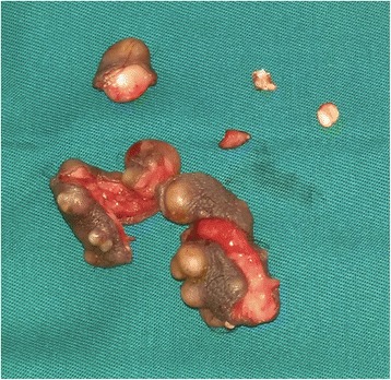
Case presentation: We report cases of two North African white patients with operated scrotal calcinosis. We describe the clinical and histological aspects as well as a pathogenic hypothesis and surgical management principles.
Conclusions: A surgical approach to scrotal calcinosis must consider the aesthetic and functional aspects postoperatively. A complete excision prevents recurrence. Psychological support is required in association with surgery because the lesions are benign and concern an intimate part of the body.
Keywords: Calcinosis; Scrotum; Surgery.
Conflict of interest statement
Ethics approval and consent to participate
No ethics committee approval was required at our institution for this case report involving a limited number of patients.
Consent for publication
Written informed consent was obtained from the patients for publication of this case report and any accompanying images. A copy of the written consents are available for review by the Editor-in-Chief of this journal.
Competing interests
The authors declare that they have no competing interests.
Publisher’s Note
Springer Nature remains neutral with regard to jurisdictional claims in published maps and institutional affiliations.
Figures


References
Publication types
MeSH terms
LinkOut - more resources
Full Text Sources
Other Literature Sources
Medical
