. 2017 Nov:211:15-21.
doi: 10.1016/j.vetmic.2017.09.020. Epub 2017 Sep 28.

Discovery of a novel swine enteric alphacoronavirus (SeACoV) in southern China

Affiliations

Discovery of a novel swine enteric alphacoronavirus (SeACoV) in southern China

Yongfei Pan et al. Vet Microbiol. 2017 Nov.

Abstract

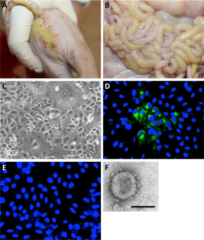
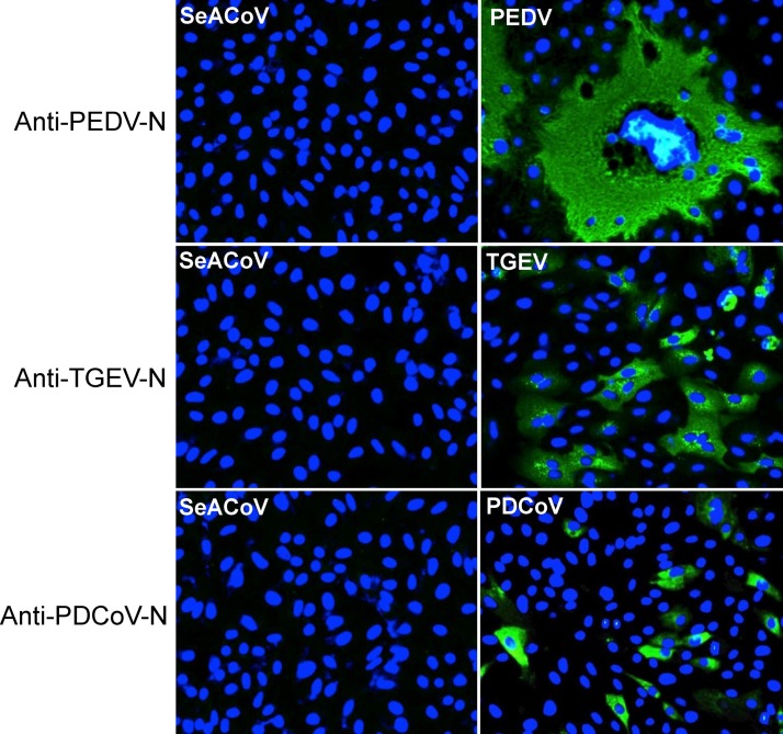
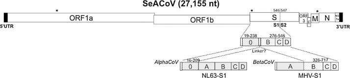
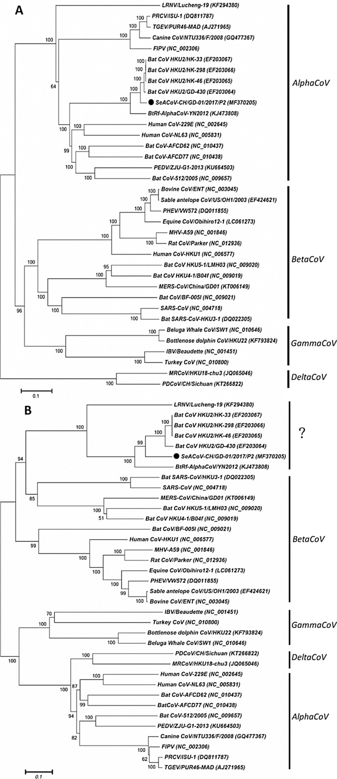
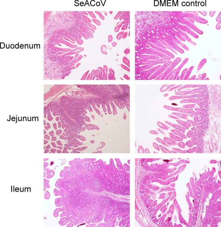
Outbreaks of diarrhea in newborn piglets without detection of transmissible gastroenteritis virus (TGEV), porcine epidemic diarrhea virus (PEDV) and porcine deltacoronavirus (PDCoV), have been recorded in a pig farm in southern China since February 2017. Isolation and propagation of the pathogen in cell culture resulted in discovery of a novel swine enteric alphacoronavirus (tentatively named SeACoV) related to the bat coronavirus HKU2 identified in the same region a decade ago. Specific fluorescence signal was detected in Vero cells infected with SeACoV by using a positive sow serum collected in the same farm, but not by using TGEV-, PEDV- or PDCoV-specific antibody. Electron microscopy observation demonstrated that the virus particle with surface projections was 100-120nm in diameter. Complete genomic sequencing and analyses of SeACoV indicated that the extreme amino-terminal domain of the SeACoV spike (S) glycoprotein structurally similar to the domain 0 of the alphacoronavirus NL63, whereas the rest part of S structurally resembles domains B to D of the betacoronavirus. The SeACoV-S domain 0 associated with enteric tropism had an extremely high variability, harboring 75-amino-acid (aa) substitutions and a 2-aa insertion, compared to that of HKU2, which is likely responsible for the extended host range or cross-species transmission. The isolated virus was infectious in pigs when inoculated orally into 3-day-old newborn piglets, leading to clinical signs of diarrhea and fecal virus shedding. These results confirmed that it is a novel swine enteric coronavirus representing the fifth porcine coronavirus.

Keywords: Bat; Cross-species transmission; Spike glycoprotein; Swine enteric alphacoronavirus (SeACoV).


Figures

Fig. 1
Fig. 1
Isolation and characterization of SeACoV. (A) A representative SeACoV-infected piglet showing clinical feature with watery feces. (B) Gross pathological lesions in the small intestine characterized by thin intestinal walls that contained moderate amounts of yellow watery feces. (C) Isolation of SeACoV-CH/GD-01/2017 in Vero cells showing CPE with syncytia formation at 48 h post-infection (hpi). (D and E) Immunofluorescence assay (IFA) result of SeACoV-infected Vero cells at 24 hpi (magnification = 200×). A positive sow serum sample (D) or a negative serum sample (E) (1:100 dilution each) collected from the disease outbreak farm was used as the first antibody followed by staining with a FITC-labeled rabbit anti-pig IgG. (F) Electron microscope image of the SeACoV/CH/GD-01 isolate (passage 5 on Vero cells) using phosphotungstic acid negative staining. Bar = 100 nm. All the results showed in panels C to F were obtained from plaque-purified virus. (For interpretation of the references to colour in this figure legend, the reader is referred to the web version of this article.)
Fig. 2
Fig. 2
IFA results of Vero cells infected with SeACoV or PEDV, Vero cells stably expressing porcine aminopeptidase N (Vero-pAPN) infected with TGEV, and LLC-PK1 cells infected with PDCoV at 48 h post-infection. SeACoV-infected Vero cells were stained with the anti-PEDV-N, anti-TGEV-N or anti-PDCoV-N monoclonal antibody, respectively (left panels). Cells infected with PEDV, TGEV or PDCoV were stained with the respective virus-specific antibody as the controls (right panels). The FITC-conjugated goat anti-mice IgG was used as the secondary Ab in IFA. Magnification = 200×.
Fig. 3
Fig. 3
Schematic diagram of the genomic structure of SeACoV and the proposed domain organization of the SeACoV spike protein S1 subunits according to the structure similarity analysis with NL63 and MHV that are both structure available. Numbers indicate amino acid positions in S glycoprotein of SeACoV, NL63 or MHV, respectively. See Supplemental Fig. S1 for the detailed sequence alignment. Nucleotide insertion/deletion at three locations (nsp3, S and M genes) in SeACoV compared to the consensus sequences of four bat-CoV HKU2 strains (GenBank accession nos. EF203064 to EF203067) are marked by “*”.
Fig. 4
Fig. 4
Phylogenetic analysis of SeACoV (GenBank accession no. MF370205) and the other representative coronaviruses based upon nucleotide sequences of the full-length genome (A) or the spike gene (B). The trees were constructed by the neighbor-joining method. Bootstrap values are indicated for each node from 1000 resamplings. The names of the viruses and strains as well as GenBank accession numbers, are shown. The black solid circle indicates the new SeACoV reported in this study.
Fig. 5
Fig. 5
Representative histological examinations of the duodenum, jejunum and ileum samples collected at 3 days post-infection from piglets inoculated with SeACoV or DMEM in the animal challenge experiment. Sections of jejunum and ileum in the SeACoV-infected group showed scattered areas of villi atrophy, whereas the section of duodenum showed mild microscopic lesions as compared to the DMEM control group.

References

    1. Akimkin V., Beer M., Blome S., Hanke D., Hoper D., Jenckel M., Pohlmann A. New chimeric porcine coronavirus in swine feces, Germany, 2012. Emerg. Infect. Dis. 2016;22:1314–1315. - PMC - PubMed
    1. Belsham G.J., Rasmussen T.B., Normann P., Vaclavek P., Strandbygaard B., Botner A. Characterization of a novel chimeric swine enteric coronavirus from diseased pigs in central Eastern Europe in 2016. Transbound. Emerg. Dis. 2016;63:595–601. - PMC - PubMed
    1. Boniotti M.B., Papetti A., Lavazza A., Alborali G., Sozzi E., Chiapponi C., Faccini S., Bonilauri P., Cordioli P., Marthaler D. Porcine epidemic diarrhea virus and discovery of a recombinant swine enteric coronavirus, Italy. Emerg. Infect. Dis. 2016;22:83–87. - PMC - PubMed
    1. de Groot R.J., Baker S.C., Baric R., Enjuanes L., Gorbalenya A.E., Holmes K.V., Perlman S., Poon L., Rottier P.J.M., Talbot P.J., Woo P.C.Y., Ziebuhr J. Coronaviridae. In: King A.M.Q., Adams M.J., Carstens E.B., Lefkowitz E.J., editors. Virus Taxonomy: Ninth Report of the International Committee on Taxonomy of Viruses. Elsevier Academic Press; London: 2011. pp. 806–828.
    1. Gong L., Li J., Zhou Q., Xu Z., Chen L., Zhang Y., Xue C., Wen Z., Cao Y. A new bat-HKU2-like coronavirus in swine, China, 2017. Emerg. Infect. Dis. 2017;23:1607–1609. - PMC - PubMed

MeSH terms