. 2018 Jan 3;26(1):320-328.
doi: 10.1016/j.ymthe.2017.09.025. Epub 2017 Oct 5.

Prostaglandin E2 Increases Lentiviral Vector Transduction Efficiency of Adult Human Hematopoietic Stem and Progenitor Cells

Affiliations

Prostaglandin E2 Increases Lentiviral Vector Transduction Efficiency of Adult Human Hematopoietic Stem and Progenitor Cells

Garrett C Heffner et al. Mol Ther. .

Abstract

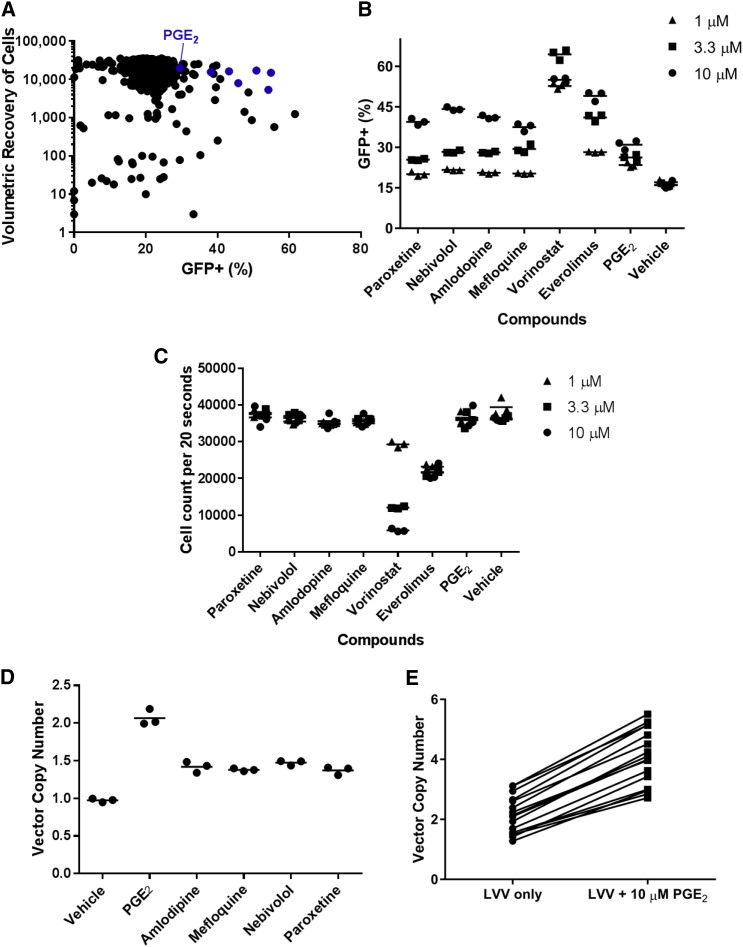
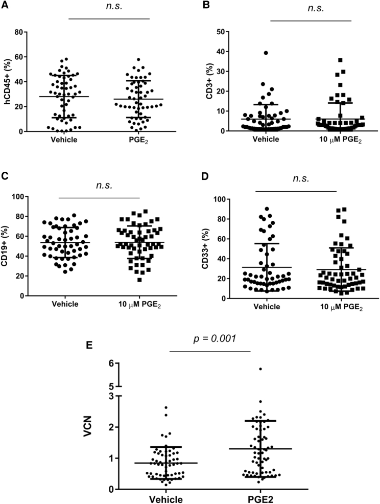
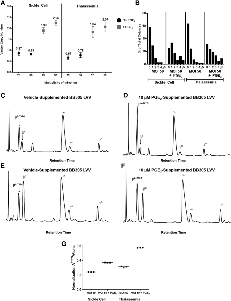
Gene therapy currently in development for hemoglobinopathies utilizes ex vivo lentiviral transduction of CD34+ hematopoietic stem and progenitor cells (HSPCs). A small-molecule screen identified prostaglandin E2 (PGE2) as a positive mediator of lentiviral transduction of CD34+ cells. Supplementation with PGE2 increased lentiviral vector (LVV) transduction of CD34+ cells approximately 2-fold compared to control transduction methods with no effect on cell viability. Transduction efficiency was consistently increased in primary CD34+ cells from multiple normal human donors and from patients with β-thalassemia or sickle cell disease. Notably, PGE2 increased transduction of repopulating human HSPCs in an immune-deficient (nonobese diabetic/severe combined immunodeficiency/interleukin-2 gamma receptor null [NSG]) xenotransplantation mouse model without evidence of in vivo toxicity, lineage bias, or a de novo bias of lentiviral integration sites. These data suggest that PGE2 improves lentiviral transduction and increases vector copy number, therefore resulting in increased transgene expression. As a result, PGE2 may be useful in clinical gene therapy applications using lentivirally modified HSPCs.

Keywords: gene therapy; hematopoietic stem cell; hemoglobinopathy; lentiviral vector; prostaglandin E(2); transduction; vector copy number.


Figures

Figure 1
Figure 1
PGE2 Enhances Transduction of CD34+ Cells with LVV (A) The results of a 780-compound small-molecule screen are depicted. Each compound is represented as a data point with the percentage of GFP+ cells indicated on the x axis and the volumetric recovery of cells, on a log scale, indicated on the y axis. Blue dots denote compounds selected for follow-up analysis. (B and C) In a follow-up experiment with 10, 3.3, or 1 μM of seven candidate compounds, the percentage of GFP+ cells (B) and the volumetric recovery of cells (C) are indicated for triplicate wells per compound per concentration of compound, as assessed at day 7 post-transduction. (D) Transduction of CD34+ cells with BB305 LVV supplemented with 10 μM candidate compound as indicated; the mean VCN is indicated for triplicate wells per compound, as assessed at day 7 post-transduction. (E) Mean VCN from CD34+ cells derived from 16 unique healthy donor cell lots, transduced with BB305 LVV supplemented with 10 μM PGE2 during the transduction step, as assessed at day 7 post-transduction. Pairwise comparisons of VCN for each cell lot, in the presence or absence of PGE2, are indicated by a line. LVV, lentiviral vector; PGE2, prostaglandin E2; VCN, vector copy number.
Figure 2
Figure 2
PGE2 Exposure during Ex Vivo Transduction Maintains huCD45+ Engraftment while Increasing VCN at a 4-Month Time Point in an NSG Xenotransplant Setting (A) Aggregate huCD45+ chimerism in bone marrow of engrafted mice at 4 months post-transplant. (B–D) Lineage analysis of huCD45+ cells in the bone marrow of engrafted mice at 4 months post-transplant, indicating the frequency of huCD45+ cells also positive for CD3 (B), CD19 (C), and CD33+ (D). (E) Aggregate mean VCN, in N = 4 experiments, in bone marrow of engrafted mice at 4 months post-transplant; 10–25 mice per group per experiment. p = 0.001 was calculated for (E) using unpaired two-tailed t test. Error bars indicate SD.
Figure 3
Figure 3
LVV Genomic Integration Profile from Xenotransplanted huCD34+ Cells Transduced in the Presence and Absence of PGE2 Bone marrow of engrafted mice from Figure 2 at 4 months post-transplant was subjected to LAM-PCR, and libraries were sequenced and aligned to reference human genome. (A) The numbers of unique reads are depicted when occurring within 30 kb upstream of a mapped transcriptional start site, within a genetic region as normalized to the transcriptional start site, or within 30 kb downstream of transcriptional termination sites. (B) Frequency of reads in locations adjacent to transcriptionally active loci, within exons, within intergenic regions, within introns, or within untranslated regions of genes. (C) Number of unique integration sites per megabase of genome, as identified in mice transplanted with cells transduced with LVV in the absence (C) or presence (D) of 10 μM PGE2. LAM-PCR, linear amplification-mediated PCR; LVV, lentiviral vector; Mb, megabase; PGE2, prostaglandin E2; TSS, transcription start site; Tx start, transcriptional start; Tx end, transcriptional end.
Figure 4
Figure 4
PGE2 Improves In Vitro Transduction of CD34+ Cells from β-Thalassemia and Sickle Cell Disease Patients (A and B) Mean VCN (A) and distribution of VCN (B) within individual colonies, after methylcellulose culture of primary CD34+ cells derived from mPB from a patient with β-thalassemia and from bone marrow from a patient with sickle cell disease, transduced with BB305 LVV, at the indicated MOI, supplemented with 10 μM PGE2 or vehicle. Data from (B) are summarized in Table 2. (A) Error bars indicate SD. (C–F) HPLC analysis of pooled BFU-E derived from methylcellulose culture of primary CD34+ cells transduced with BB305 LVV and supplemented with vehicle (C and E) or 10 μM PGE2 (D and F). Data from triplicate HPLC samples for (C–F) are summarized in (G). (G) Data from HPLC samples (C–F), performed in triplicate, are summarized. BFU-E, burst-forming unit-erythroid; HPLC, high-performance liquid chromatography; LVV, lentiviral vector; mBP, mobilized peripheral blood; MOI, multiplicity of infection; PGE2, prostaglandin E2; VCN, vector copy number.

Similar articles

Cited by

References

    1. Biffi A., Montini E., Lorioli L., Cesani M., Fumagalli F., Plati T., Baldoli C., Martino S., Calabria A., Canale S. Lentiviral hematopoietic stem cell gene therapy benefits metachromatic leukodystrophy. Science. 2013;341:1233158. - PubMed
    1. Cavazzana-Calvo M., Payen E., Negre O., Wang G., Hehir K., Fusil F., Down J., Denaro M., Brady T., Westerman K. Transfusion independence and HMGA2 activation after gene therapy of human β-thalassaemia. Nature. 2010;467:318–322. - PMC - PubMed
    1. Aiuti A., Biasco L., Scaramuzza S., Ferrua F., Cicalese M.P., Baricordi C., Dionisio F., Calabria A., Giannelli S., Castiello M.C. Lentiviral hematopoietic stem cell gene therapy in patients with Wiskott-Aldrich syndrome. Science. 2013;341:1233151. - PMC - PubMed
    1. Scaramuzza S., Biasco L., Ripamonti A., Castiello M.C., Loperfido M., Draghici E., Hernandez R.J., Benedicenti F., Radrizzani M., Salomoni M. Preclinical safety and efficacy of human CD34(+) cells transduced with lentiviral vector for the treatment of Wiskott-Aldrich syndrome. Mol. Ther. 2013;21:175–184. - PMC - PubMed
    1. Sessa M., Lorioli L., Fumagalli F., Acquati S., Redaelli D., Baldoli C., Canale S., Lopez I.D., Morena F., Calabria A. Lentiviral haemopoietic stem-cell gene therapy in early-onset metachromatic leukodystrophy: an ad-hoc analysis of a non-randomised, open-label, phase 1/2 trial. Lancet. 2016;388:476–487. - PubMed

Publication types

MeSH terms