

Atypical Callosal Morphology in Children with Speech Sound Disorder
- PMID: 29102664
- PMCID: PMC5709239
- DOI: 10.1016/j.neuroscience.2017.10.039
Atypical Callosal Morphology in Children with Speech Sound Disorder
Abstract
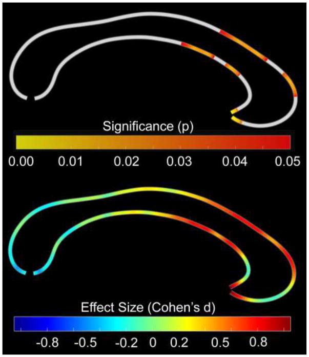
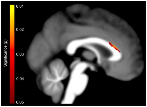
Speech sound disorder (SSD) is common, yet its neurobiology is poorly understood. Recent studies indicate atypical structural and functional anomalies either in one hemisphere or both hemispheres, which might be accompanied by alterations in inter-hemispheric connectivity. Indeed, abnormalities of the corpus callosum - the main fiber tract connecting the two hemispheres - have been linked to speech and language deficits in associated disorders, such as stuttering, dyslexia, aphasia, etc. However, there is a dearth of studies examining the corpus callosum in SSD. Here, we investigated whether a sample of 18 children with SSD differed in callosal morphology from 18 typically developing children carefully matched for age. Significantly reduced dimensions of the corpus callosum, particularly in the callosal anterior third, were observed in children with SSD. These findings indicating pronounced callosal aberrations in SSD make an important contribution to an understudied field of research and may suggest that SSD is accompanied by atypical lateralization of speech and language function.
Keywords: anterior third; brain; corpus callosum; development; language; magnetic resonance imaging.
Copyright © 2017 IBRO. Published by Elsevier Ltd. All rights reserved.
Figures
References
-
- Achiron R, Achiron A. Development of the human fetal corpus callosum: a high-resolution, cross-sectional sonographic study. Ultrasound Obstet Gynecol. 2001;18:343–347. - PubMed
-
- Anastasopoulou S, Kurth F, Luders E, Savic I. Generalized epilepsy syndromes and callosal thickness: Differential effects between patients with juvenile myoclonic epilepsy and those with generalized tonic-clonic seizures alone. Epilepsy Res. 2016;129:74–78. - PubMed
-
- Ashburner J. A fast diffeomorphic image registration algorithm. Neuroimage. 2007;38:95–113. - PubMed
-
- Beitchman JH, Wilson B, Johnson CJ, Atkinson L, Young A, Adlaf E, Escobar M, Douglas L. Fourteen-year follow-up of speech/language-impaired and control children: psychiatric outcome. J Am Acad Child Adolesc Psychiatry. 2001;40:75–82. - PubMed
MeSH terms
Grants and funding
LinkOut - more resources
Full Text Sources
Other Literature Sources
