Delayed ischemic stroke due to stent marker band occlusion after stent-assisted coiling
- PMID: 29102968
- PMCID: PMC5747672
- DOI: 10.1136/bcr-2017-013364
Delayed ischemic stroke due to stent marker band occlusion after stent-assisted coiling
Abstract
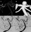
A middle-aged patient with an internal carotid-posterior communicating artery aneurysm and basilar artery tip aneurysm was treated by stent-assisted coiling. One ischemic infarction and two transient ischemic attacks occurred with the same symptoms (inability to walk unassisted and tendency to fall to the left) during the first 2 years post-treatment. The ischemic infarction was found in the right side of the pons, consistent with the vascular territory of the stent-containing vessel. The cause of the delayed ischemic stroke was investigated on DSA and cone beam CT, which revealed that the proximal end of the stent, one marker band, was just covering a small perforating artery of the basilar artery trunk. The present case suggests that marker band occlusion can induce delayed ischemic stroke. To prevent this complication, it is important to evaluate the perforating vessels preoperatively and carefully deploy a stent for the marker band to avoid occlusion of large perforating vessels. Post-treatment evaluation is also important because dual antiplatelet therapy will be required for a longer period if an artery is occluded by a marker band.
Keywords: aneurysm; platelets; stent; stroke.
© BMJ Publishing Group Ltd (unless otherwise stated in the text of the article) 2017. All rights reserved. No commercial use is permitted unless otherwise expressly granted.
Conflict of interest statement
Competing interests: None declared.
Figures


Similar articles
-
Delayed ischemic stroke due to stent marker band occlusion after stent-assisted coiling.J Neurointerv Surg. 2018 Aug;10(8):e20. doi: 10.1136/neurintsurg-2017-013364.rep. Epub 2018 Jun 16. J Neurointerv Surg. 2018. PMID: 29909378
-
Very late ischemic complications in flow-diverter stents: a retrospective analysis of a single-center series.J Neurosurg. 2016 Oct;125(4):929-935. doi: 10.3171/2015.10.JNS15703. Epub 2016 Jan 29. J Neurosurg. 2016. PMID: 26824382
-
Midterm results of T-stent-assisted coiling of wide-necked and complex intracranial bifurcation aneurysms using low-profile stents.J Neurosurg. 2017 Dec;127(6):1288-1296. doi: 10.3171/2016.9.JNS161909. Epub 2017 Jan 6. J Neurosurg. 2017. PMID: 28059656
-
Delayed coil migration from a small wide-necked aneurysm after stent-assisted embolization: case report and literature review.Neuroradiology. 2006 May;48(5):333-7. doi: 10.1007/s00234-005-0044-1. Epub 2006 Apr 6. Neuroradiology. 2006. PMID: 16598480 Review.
-
Nonoverlapping Y-configuration stenting technique with dual closed-cell stents in wide-neck basilar tip aneurysms.Neurosurgery. 2012 Jun;70(2 Suppl Operative):244-9. doi: 10.1227/NEU.0b013e31823bcdc5. Neurosurgery. 2012. PMID: 21993186 Review.
References
Publication types
MeSH terms
LinkOut - more resources
Full Text Sources
Other Literature Sources
Medical
