. 2018 Apr;125(4):537-548.
doi: 10.1016/j.ophtha.2017.09.028. Epub 2017 Nov 2.

Consensus Definition for Atrophy Associated with Age-Related Macular Degeneration on OCT: Classification of Atrophy Report 3

Affiliations

Consensus Definition for Atrophy Associated with Age-Related Macular Degeneration on OCT: Classification of Atrophy Report 3

Srinivas R Sadda et al. Ophthalmology. 2018 Apr.

Erratum in

  • Corrigendum.
    [No authors listed] [No authors listed] Ophthalmology. 2019 Jan;126(1):177. doi: 10.1016/j.ophtha.2018.10.038. Ophthalmology. 2019. PMID: 30577915 No abstract available.

Abstract

Purpose: To develop consensus terminology and criteria for defining atrophy based on OCT findings in the setting of age-related macular degeneration (AMD).

Design: Consensus meeting.

Participants: Panel of retina specialists, image reading center experts, retinal histologists, and optics engineers.

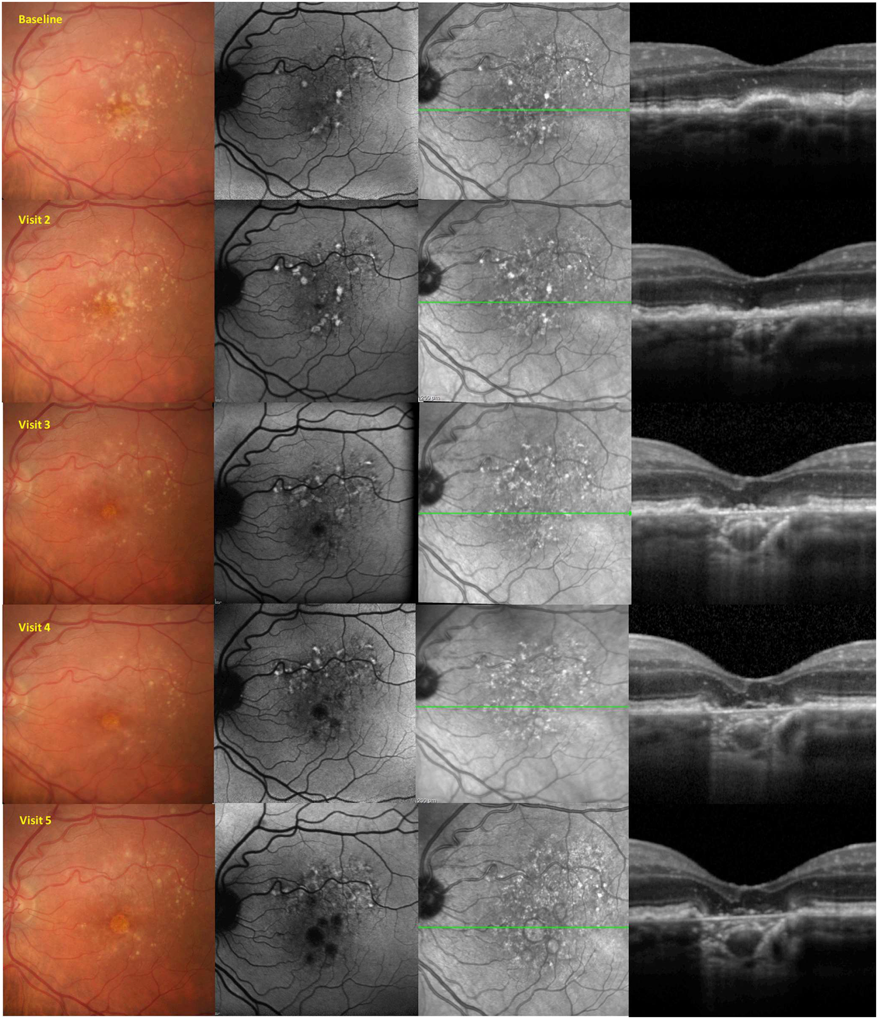
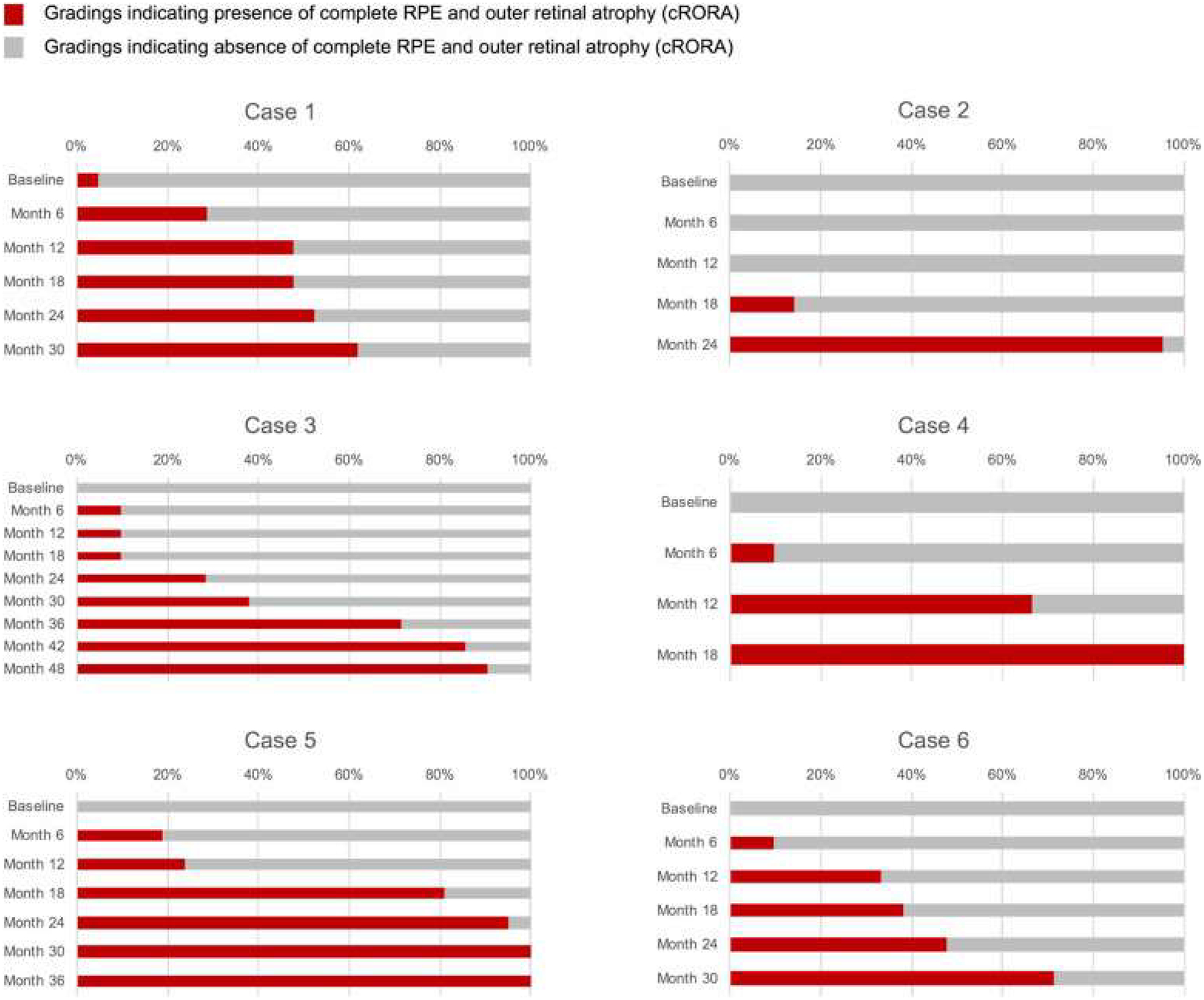
Methods: As part of the Classification of Atrophy Meetings (CAM) program, an international group of experts surveyed the existing literature, performed a masked analysis of longitudinal multimodal imaging for a series of eyes with AMD, and reviewed the results of this analysis to define areas of agreement and disagreement. Through consensus discussions at 3 meetings over 12 months, a classification system based on OCT was proposed for atrophy secondary to AMD. Specific criteria were defined to establish the presence of atrophy.

Main outcome measures: A consensus classification system for atrophy and OCT-based criteria to identify atrophy.

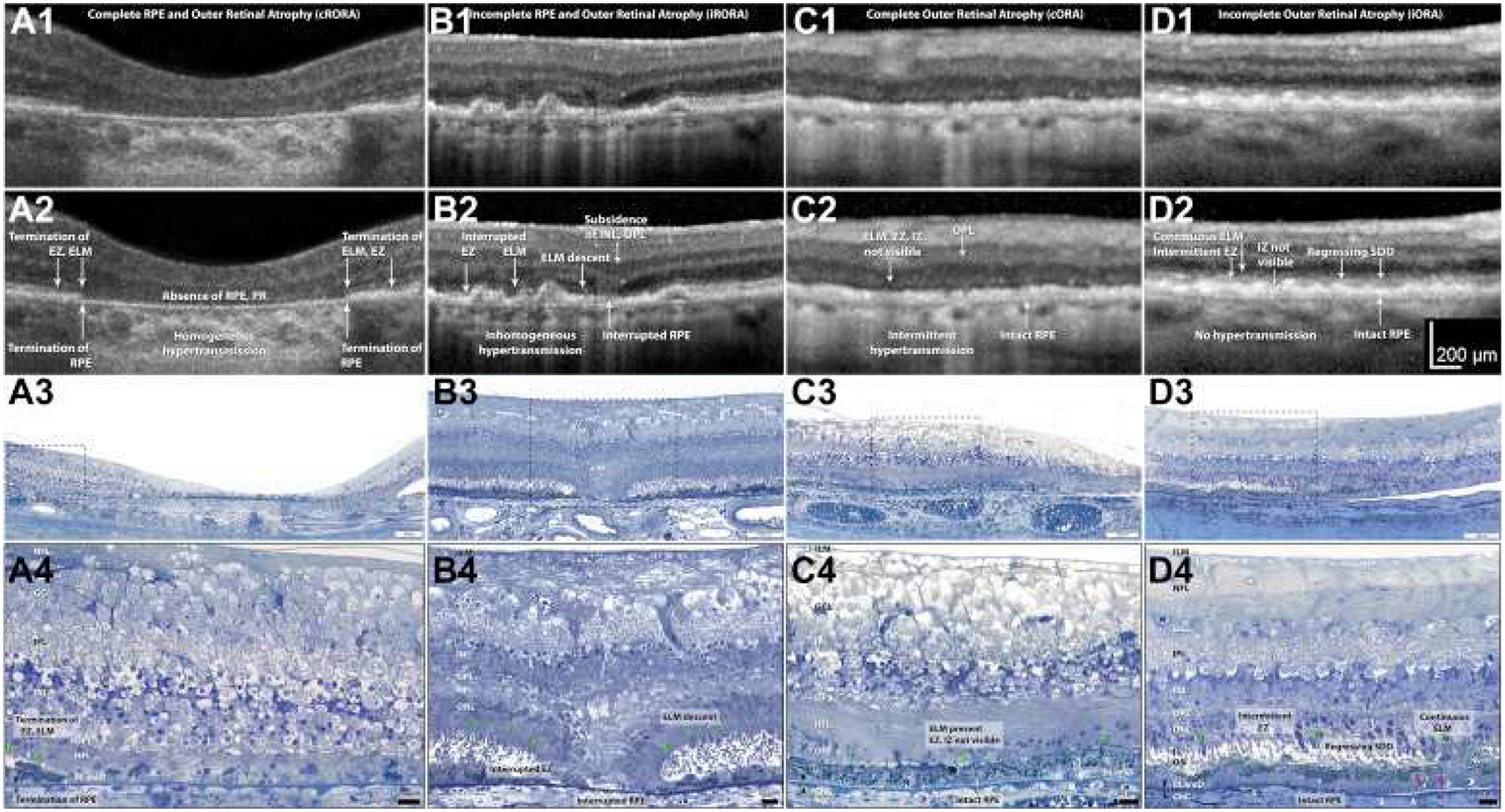
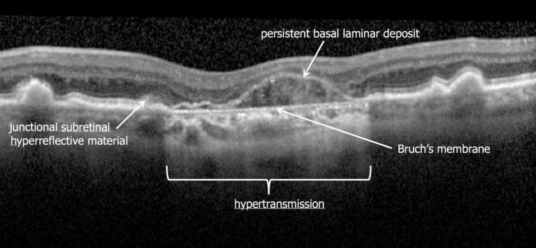
Results: OCT was proposed as the reference standard or base imaging method to diagnose and stage atrophy. Other methods, including fundus autofluorescence, near-infrared reflectance, and color imaging, provided complementary and confirmatory information. Recognizing that photoreceptor atrophy can occur without retinal pigment epithelium (RPE) atrophy and that atrophy can undergo an evolution of different stages, 4 terms and histologic candidates were proposed: complete RPE and outer retinal atrophy (cRORA), incomplete RPE and outer retinal atrophy, complete outer retinal atrophy, and incomplete outer retinal atrophy. Specific OCT criteria to diagnose cRORA were proposed: (1) a region of hypertransmission of at least 250 μm in diameter, (2) a zone of attenuation or disruption of the RPE of at least 250 μm in diameter, (3) evidence of overlying photoreceptor degeneration, and (4) absence of scrolled RPE or other signs of an RPE tear.

Conclusions: A classification system and criteria for OCT-defined atrophy in the setting of AMD has been proposed based on an international consensus. This classification is a more complete representation of changes that occur in AMD than can be detected using color fundus photography alone. Longitudinal information is required to validate the implied risk of vision loss associated with these terms. This system will enable such future studies to be undertaken using consistent definitions.


Figures

Figure 1.
Figure 1.
Example of a premeeting exercise distributed to the Classification of Atrophy Meetings participants. From this longitudinal multimodal imaging dataset, participants were asked to define the earliest time point at which complete atrophy and complete retinal pigment epithelium and outer retinal atrophy were present.
Figure 2.
Figure 2.
Exemplar OCT B-scans and representative corresponding histologic images demonstrating the 4 terms for atrophy defined by the Classification of Atrophy Meetings. Rows 1 and 2 show B-scans of each atrophy phenotype, without and with annotations, respectively. Rows 3 and 4 show histologic images of different eyes (i.e., not those in the B-scans) at medium and high magnification, respectively. Nomenclature and annotations are given below. OCT scans and photomicrographs of normal retinas are available in Figures S1 and S2 (available at www.aaojoumal.org), respectively; methods for histologic examination and photomicroscopy are in Figure S2 (available at www.aaojoumal.org). A, Images obtained from an 83-year-old white woman. A1, A2, Complete retinal pigment epithelium (RPE) and outer retinal atrophy (cRORA) is defined by a zone of homogeneous choroidal hypertransmission and absence of the RPE band measuring 250 μm or more with overlying outer retinal thinning and loss of photoreceptors (PR). No signs of an RPE tear are evident. Terminations of the external limiting membrane (ELM), ellipsoid zone (EZ), and RPE are indicated. A3, A4, In a region of fovea and parafovea measuring 1 mm or more in diameter, RPE cells are absent, with some dissociated RPE cells remaining. Complete loss of photoreceptors is shown, that is, the outer nuclear layer (ONL), ELM, inner segment (IS), and outer segment (OS) are not apparent. The Henle fiber layer (HFL) in the atrophic zone consists primarily of Muller cell fibers that do not make an ELM among themselves. B, Images obtained from an 87-year-old white man. B1, B2, For incomplete RPE and outer retinal atrophy (iRORA), some hypertransmission is evident but is discontinuous; the RPE band is present but irregular or interrupted. Interrupted ELM and EZ evidences photoreceptor degeneration. The inner nuclear layer (INL) and outer plexiform layer (OPL) exhibit subsidence. Criteria for cRORA are not met. B3, B4, The ELM descends in 2 curved lines (green arrowheads). The ONL, HFL, OPL, and INL subside in parallel to the ELM, creating a funnel. The RPE layer is interrupted. The IS are short, and the OS are absent near the ELM descent. C, Images from an 88-year-old-white woman. C1, C2, Complete outer retinal atrophy (cORA) is defined by continuous nonvisibility of the EZ and interdigitation zone (IZ) and severe thinning of the outer retina, in the setting of an intact RPE band. Hypertransmission associated with RPE degeneration is intermittent. C3, C4, The RPE layer largely is intact, although individual cells are dysmorphic. The OS and IS are completely absent. The ONL is thin or interrupted, with several clumps of RPE-derived material, and the ELM is intact. The HFL contains fibers of Müller cells and some photoreceptors. D, Images from an 85-year-old white woman. D1, D2. Incomplete outer retinal atrophy (iORA) demonstrating continuous ELM and detectable EZ disruption in the setting of regressing subretinal drusenoid deposits (SDD), with detectable thinning of the outer retina, an intact RPE band, and no hypertransmission. D3, D4, The RPE layer largely is intact, although individual cells are dysmorphic. Some isolated subretinal drusenoid deposits (Regressing SDD) are present above the RPE, but OS and IS remain. The ONL is depopulated with intact ELM and pale-staining reactive Muller processes between the photoreceptor nuclei. Photoreceptors span layers OPL, HFL, ONL, IS, and OS. Muller cells span the internal limiting membrane (ILM) to ELM. In naming the OPL and HFL, we use the OCT designations while recognizing that a commonly used neurobiological nomenclature divides the OPL into 2 sublayers (photoreceptor synaptic terminals and bipolar or horizontal cell dendrites) and combines these with the HFL into 1 “OPL.” White arrowheads = calcified Bruch’s membrane; pink arrowheads = basal mounds; X = artifactual wrinkle; black arrowheads = Bruch’s membrane; green arrowheads = ELM. BLamD = basal laminar deposit; ChC = choriocapillaris; GCL = ganglion cell layer; IPL = inner plexiform layer; NFL = nerve fiber layer. Figure prepared by M. Li and J. D. Messinger.
Figure 3.
Figure 3.
Example of a premeeting exercise distributed to the Classification of Atrophy Meetings (CAM) participants. Arrows were used to mark specific features on images (in this case an OCT B-scan) for which participants were asked to provide specific terms or labels. Consensus labels for 4 specific features after discussions at CAM are shown in this illustration. Note the thinning of the outer retina over the persistent basal laminar deposit band.,
Figure 4.
Figure 4.
Cumulative frequency of Classification of Atrophy Meetings (CAM) participants indicating that the complete retinal pigment epithelium (RPE) and outer retinal atrophy (cRORA) had been achieved for 6 patients with multimodal imaging. For each patient, each row represents 1 visit or time point, with the baseline visit at the top and the final visit at the bottom. The length of the bar indicates the percentage of CAM participants who deemed the cRORA end point was present on the OCT images for that visit. Note for patient 2, only 6 months elapsed between when at least 1 CAM participant determined that cRORA was present and the vast majority believed that it was present. In other patients, the interval was considerably longer, highlighting initial disagreement among participants regarding the criteria used for establishing presence of atrophy on OCT.
Figure 5.
Figure 5.
Montage of the OCT images for the 6 patients shown in Figure 4 at the first time point at which at least 60% of Classification of Atrophy Meetings participants determined that complete retinal pigment epithelium (RPE) and outer retinal atrophy (cRORA) was present. Patients 2 and 5 most clearly illustrate the typical features of cRORA, with choroidal hypertransmission and attenuation of the RPE band with thinning of the overlying retina in a region exceeding 250 μm in diameter.

References

    1. Ferris FL 3rd, Wilkinson CP, Bird A, et al. Clinical classification of age-related macular degeneration. Ophthalmology. 2013;120:844–851. - PMC - PubMed
    1. Holz FG, Strauss EC, Schmitz-Valckenberg S, van Lookeren Campagne M. Geographic atrophy: clinical features and potential therapeutic approaches. Ophthalmology. 2014;121:1079–1091. - PubMed
    1. Gass JD. Stereoscopic Atlas of Macular Diseases. 1st ed. St. Louis: The C. V. Mosby Company; 1970.
    1. Schmitz-Valckenberg S, Sadda S, Staurenghi G, et al. Geographic atrophy: semantic considerations and literature review. Retina. 2016;36:2250–2264. - PMC - PubMed
    1. Haab O Erkrankungen der Macula Lutea. Centralblat Augenheilkd. 1885;9:384–391.

Publication types