Review
. 2018 Feb;28(2):113-127.
doi: 10.1016/j.tcb.2017.10.001. Epub 2017 Nov 2.

Reading m6A in the Transcriptome: m6A-Binding Proteins

Affiliations
Review

Reading m6A in the Transcriptome: m6A-Binding Proteins

Deepak P Patil et al. Trends Cell Biol. 2018 Feb.

Abstract

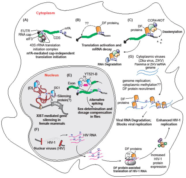
N6-Methyladenosine (m6A) is the most prevalent post-transcriptional modification of eukaryotic mRNA and long noncoding RNA. m6A mediates its effects primarily by recruiting proteins, including the multiprotein eukaryotic initiation factor 3 complex and a set of proteins that contain the YTH domain. Here we describe the mechanisms by which YTH domain-containing proteins bind m6A and influence the fate of m6A-containing RNA in mammalian cells. We discuss the diverse, and occasionally contradictory, functions ascribed to these proteins and the emerging concepts that are influencing our understanding of these proteins and their effects on the epitranscriptome.

Keywords: N(6)-methyl adenosine; RNA metabolism; YTH proteins; m(6)A modification; splicing; translational regulation.


Figures

Figure 1
Figure 1. Domain structure and functions of human YTH proteins
Schematic representation of domains and disordered regions of human YTH proteins: DF1, DF2, DF3, DC1, and DC2. DF1, DF2, and DF3 together make the YTHDF (DF) family. DC2 and the DF family proteins have a C-terminal YTH domain (pink), while DC1 has an internal YTH domain. All the proteins have low-complexity disordered regions (green). Disordered regions were identified using D2P2 database tools [80]. Additional domains in DC2 include R3H, DEXDc, ankyrin repeats (ANK), HELICc, HA2 and OB-fold domains. Protein length (in amino acids) is indicated next to each protein schematic. Functions of the human YTH proteins along with their alias names and major cellular localization are also indicated in a table (bottom panel).
Figure 2
Figure 2. Human YTHDF proteins are paralogs of each other
Shown is a species-aware phylogenetic gene tree of YTHDF family genes across the indicated organisms or group of organisms. This tree was constructed using the Ensembl Gene orthology and paralogy prediction pipeline [81]. Gene duplication events ( formula image
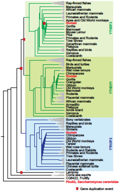
) and YTHDF1, YTHDF2, and YTHDF3 gene clades are also indicated.
Figure 3
Figure 3. A hydrophobic aromatic cage in the YTH domain recognizes m6A
Shown here is the organization of m6A (blue) in the WWY-type aromatic cage (pink) of Z. rouxii MRB1 YTH domain. A tyrosine (Y260) and tryptophan (W200) residues in the protein sandwich the 6-methyladenine group. This positions the methyl group pointing to another tryptophan (W254) which forms the base of the cage. Notably, the purine ring of the m6A residue is not involved in this hydrophobic interaction. The amino acid positions in the domain are highly conserved across species. The YTHDF family proteins of proteins have a WWW cage, and YTHDC1 and YTHDC2 have a WWL-type cage.
Figure 4
Figure 4. YTH domains share a conserved mode of binding m6A RNA
Amino acid sequence alignment of the YTH domains from the indicated organisms is shown here. Secondary structural elements in the YTH domain from Z. rouxii MRB1 (PDB: 4U8T) are shown above the sequence alignment. Amino acid positions in individual protein are also indicated next to the protein names. Amino acids participating in recognition of different parts of m6A RNA are indicated with filled shapes below the sequence alignment. Above the alignments: TT, strict β-turns; arrows, β-strands; coils, α-helices. In the alignments: red shade with white characters, identical amino acids; red characters, similar amino acids; blue frame, marks block of sequence with similarity. Below the alignments: ( formula image
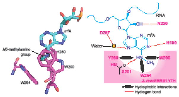
) Residues making contact with bases other than m6A in Z. rouxii MRB1; (●) Residues making contact with the m6A base in Z. rouxii MRB1; (π) π-stacking Y205 in Z. rouxii MRB1 with G upstream of the m6A nucleotide; not conserved in human DC2; (π) π-stacking R296 in Z. rouxii MRB1 with the C on the 3′ side of the m6A nucleotide; ( formula image
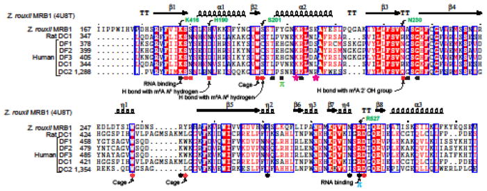
) R1318 and R1322 in human DC2 form an increased positive surface charge than in human DC1. Amino acids involved in cage formation, RNA binding, hydrogen bond (H bond) formation are also indicated.
Figure 5
Figure 5. Recognition of m6A in human MAT2A mRNA and rRNAs
(a) YTH domain proteins recognize m6A in CAG motif. 3′UTR of MAT2A mRNA contains m6A in an evolutionarily conserved CAG motif in U6-like hairpin loops. Shown here is a gene model of MAT2A 3′UTR (dark blue), m6A miCLIP tag profile (purple track) and iCLIP tag-based binding profiles of human DF1, DF2, DF3, DC1, and DC2 proteins. Dotted boxes mark the overlapping DF1, DF2, DF3 and DC1 peaks and the methylated regions on MAT2A 3′UTR. The green shaded portion on the hairpin loops marks the N6-methyladenosine containing CAG motif (zoomed-in view). (b) YTH proteins do not bind m6A on rRNAs. The highly prevalent m6A-containing 28S and 18S rRNAs each have one m6A modification in a AAC sequence motif. Shown is the location of m6A in 28S rRNA (left) and 18S rRNA (right), along with m6A peaks (miCLIP, purple), and the iCLIP binding profile of endogenous human YTH proteins. The absence of a peak in the YTH iCLIP reads show that none of the five YTH proteins bind at the m6A site on either 28S or 18S rRNA.
Figure 6, Key Figure
Figure 6, Key Figure. Multiple functions of YTH proteins
(a) Cap-independent translation mediated by m6A and eIF3. A subset of transcripts contain m6As in their 5′UTR that can be recognized by eIF3. Binding of eIF3 recruits the 43S translation initiation complex leading to cap-independent translation. (b) YTHDF proteins regulate translation of transcripts by binding to m6A in the 3′UTR and recruiting eIF3. It is proposed that the mRNA circularizes to bring eIF3 in proximity to the cap in order to initiate translation. (c) YTHDF proteins promote mRNA decay. The C-terminal YTH domain binds m6A on transcripts and the N-terminal disordered region recruits CNOT1, a component of the CCR4-NOT complex. Following deadenylation by CCR4-NOT1 transcripts are decapped and degraded. (d) YTHDC1 is necessary for X-chromosome inactivation (XCI). XIST is a heavily methylated ncRNA bound by DC1. In the absence of m6A or DC1 X-chromosome inactivation is impaired. (e) YTHDC1 (YT-521B in Drosophila) binding to m6A is necessary for X chromosome dosage compensation and sex determination in flies. In female flies, the introns flanking the second exon of the sex-lethal (Sxl) transcript contain m6A and binding of DC1 promotes skipping of the second exon and production of full-length Sxl protein. (f) Nuclear replicating viruses, such as HIV, have highly conserved m6A sites that actively recruit DF proteins to promote translation of viral proteins. (G) Cytoplasmic replicating viruses, such as Zika virus, are methylated in the cytoplasm through an unknown METTL3-dependent mechanism and bound by the DF proteins. Binding of DF proteins to cytoplasmic virus transcripts confines the transcripts to lipid droplets in liver cells and inhibits viral replication.

References

    1. Perry RP, Kelley DE. Existence of methylated messenger RNA in mouse L cells. Cell. 1974;1(1):37–42.
    1. Desrosiers R, et al. Identification of methylated nucleosides in messenger RNA from Novikoff hepatoma cells. Proc Natl Acad Sci U S A. 1974;71(10):3971–5. - PMC - PubMed
    1. Clancy MJ, et al. Induction of sporulation in Saccharomyces cerevisiae leads to the formation of N6-methyladenosine in mRNA: a potential mechanism for the activity of the IME4 gene. Nucleic Acids Res. 2002;30(20):4509–18. - PMC - PubMed
    1. Zhong S, et al. MTA is an Arabidopsis messenger RNA adenosine methylase and interacts with a homolog of a sex-specific splicing factor. Plant Cell. 2008;20(5):1278–88. - PMC - PubMed
    1. Meyer KD, et al. Comprehensive analysis of mRNA methylation reveals enrichment in 3′ UTRs and near stop codons. Cell. 2012;149(7):1635–46. - PMC - PubMed

Publication types