

Fluorescence Microscopy Imaging Calibration for Quantifying Nanocarrier Binding to Cells During Shear Flow Exposure
- PMID: 29104516
- PMCID: PMC5665578
- DOI: 10.1166/jbn.2017.2392
Fluorescence Microscopy Imaging Calibration for Quantifying Nanocarrier Binding to Cells During Shear Flow Exposure
Abstract
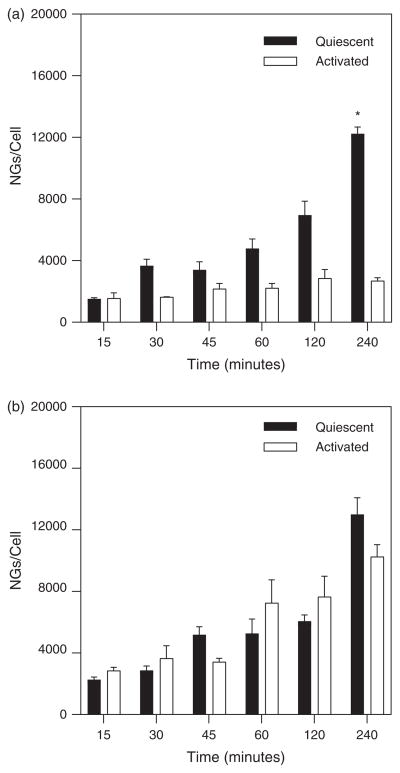
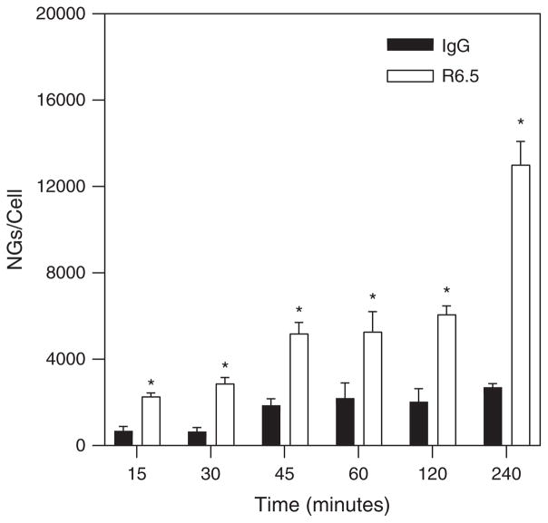
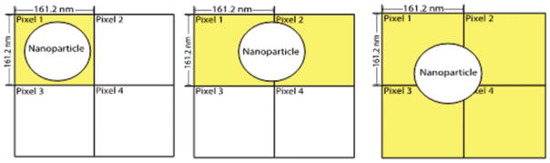
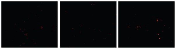
Targeted drug delivery is a fast growing industry in healthcare and technologies are being developed for applications utilizing nanocarriers as vehicles for drug transport. As the size scale of these particles becomes further reduced, advanced fluorescence microscopy and image analysis techniques become increasingly important for facilitating our understanding of nanocarrier binding and avidity, thereby establishing the basis for nanocarrier design optimization. While there is a significant body of published work using nanocarriers in vitro and in vivo, the advent of smaller particles that have typically been studied (~500 nm) limits the ability to attain quantitative measurements of nanocarrier binding dynamics since image acquisition and analysis methods are restricted by microscopy pixel size. This work demonstrates the use of a novel calibration technique based on radioisotope counting and fluorescence imaging for enabling quantitative determination of nanocarrier binding dynamics. The technique is then applied to assess the temporal profile of endothelial cell binding of two antibody targeted nanocarrier types in the presence of fluid shear stress. Results are provided for binding of nanoparticles smaller than a microscopy image pixel.
Keywords: Binding; Calibration; Fluorescence Microscopy; Image Analysis; Nanocarrier; Nanogel; Quantitative Binding; Shear Stress; Targeted Drug Delivery.
Figures
References
-
- Ding BS, Dziubla T, Shuvaev VV, Muro S, Muzykantov VR. Mol Interv. 2006;6:98. - PubMed
-
- Mastrobattista E, Storm G, van Bloois L, Reszka R, Bloemen PG, Crommelin DJ, Henricks PA. Biochim Biophys Acta. 1999;1419:353. - PubMed
-
- Weller GER, Villanueva FS, Klibanov AL, Wagner WR. Ann Biomed Eng. 2002;30:1012. - PubMed
MeSH terms
Substances
Grants and funding
LinkOut - more resources
Full Text Sources
Other Literature Sources
Research Materials