. 2017 Nov;14(5):4194-4200.
doi: 10.3892/etm.2017.5090. Epub 2017 Sep 1.

Upregulation of T-cadherin suppresses cell proliferation, migration and invasion of gastric cancer in vitro

Affiliations

Upregulation of T-cadherin suppresses cell proliferation, migration and invasion of gastric cancer in vitro

Jianqing Lin et al. Exp Ther Med. 2017 Nov.

Abstract

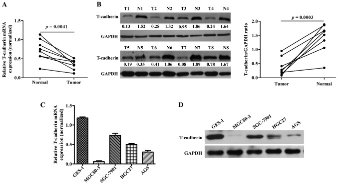
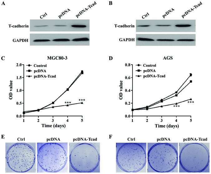
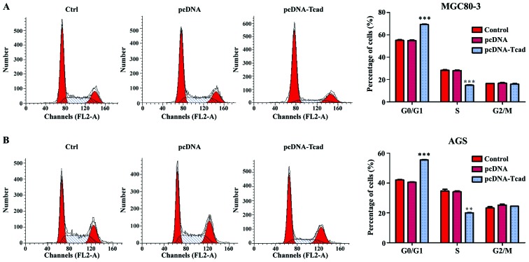
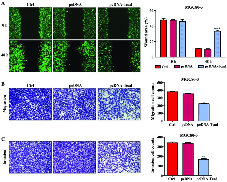
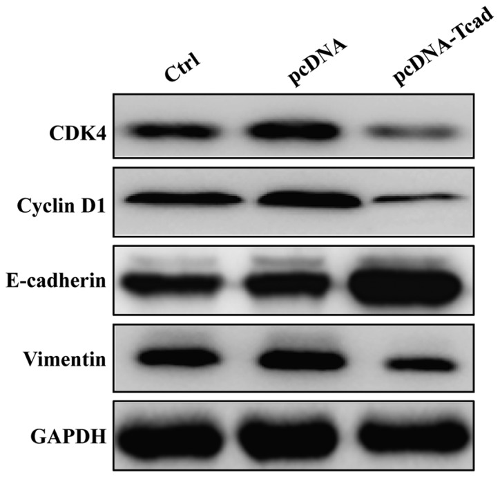
As a unique member of the cadherin superfamily, T-cadherin (T-cad) has been demonstrated to be associated with gastric cancer (GC) prognosis. To elucidate the function of T-cad in GC in vitro, the present study firstly examined T-cad protein expression in normal and gastric cancer tissues and cell lines, and it was demonstrated to be significantly downregulated in gastric cancer samples compared with normal samples. Control and T-cad expression vectors were then transfected into the MGC8-03 and AGS GC cell lines. Utilizing MTT, clonogenic, flow cytometry, wound healing and Transwell invasion assays in addition to Western blotting, the present study demonstrated that the overexpression of T-cad suppressed GC cell growth and colony formation via cell cycle arrest at the G0/G1 phase via downregulating the expression of cyclin dependent kinase 4 and Cyclin D1. In addition, overexpression of T-cad significantly inhibited GC cell migration and invasion by increasing E-cadherin and decreasing Vimentin expression. These findings suggest T-cad may be important in GC cell proliferation and metastasis and serve as a promising target for the treatment of GC in the future.

Keywords: T-cadherin; cell cycle; cell proliferation; gastric cancer; invasion; migration.


Figures

Figure 1.
Figure 1.
T-cadherin is downregulated in GC tissues and cell lines. The mRNA and protein levels of T-cadherin in GC tissues and adjacent noncancerous tissues were determined using (A) qRT-PCR and (B) western blotting analyses, respectively. The (C) qRT-PCR and (D) western blotting were used to measure the mRNA and protein levels of T-cadherin in GC cell lines (MGC80-3, SGC-7901, HGC27 and AGS) and normal GES-1 cell line, respectively. Quantitative data were expressed as mean ± standard deviation of three independent experiments. GC, gastric cancer; N, adjacent noncancerous tissues; T, GC tissues; GAPDH serves as an internal control.
Figure 2.
Figure 2.
T-cadherin inhibits GC cell proliferation. Overexpression of T-cadherin protein in transfected cell lines, (A) MGC80-3 and (B) AGS was confirmed by western blotting. MTT assay was used to measure cell viability in (C) MGC80-3 and (D) AGS after transfection with empty vector pcDNA or pcDNA-T-cadherin (pcDNA-Tcad). These data are shown as the mean ± standard deviation of three independent experiments. The size and number of colonies formed in (E) MGC80-3 and (F) AGS cells recorded under a light microscope. The representative pictures shown are from one of three independent experiments. ***P<0.001 vs. control or empty vector pcDNA.
Figure 3.
Figure 3.
T-cadherin arrested cell cycle at G0/G1 phase in GC cells. Flow cytometry was used to analyze cell cycle distribution in empty vector pcDNA or pcDNA-T-cadherin (pcDNA-Tcad)-transfected (A) MGC80-3 and (B) AGS cells. Left panel shows representative dot plots and right panels show the quantitative analysis. These data are shown as the mean ± standard deviation of three independent experiments. **P<0.01, ***P<0.001 vs. control or empty vector pcDNA.
Figure 4.
Figure 4.
T-cadherin suppressed GC cell motility, migration and invasion. (A) Wound-healing assay was used to evaluate motility of cells transfected with control, empty vector pcDNA, pcDNA-Tcad. The wound area was visualized and calculated at 1 and 48 h. (B) Cell migration transfected with control, empty vector pcDNA, pcDNA-Tcad was examined by transwell assay. (C) The cell invasive ability transected with control, empty vector pcDNA, pcDNA-Tcad was determined by Matrigel cell culture chambers. These data are shown as the mean ± standard deviation of three independent experiments. *P<0.05, **P<0.01, ***P<0.001 vs. control or empty vector pcDNA.
Figure 5.
Figure 5.
Effect of T-cadherin on markers associated with cell cycle progression and metastatic potential in GC. Western blotting was used to analyze the expression of CDK4, Cyclin D1, E-cadherin, Vimentin and MMP2 in MGC80-3 cells following control, empty vector pcDNA, pcDNA-Tcad transfection, respectively. GAPDH serves as an internal control.

References

    1. Piazuelo MB, Correa P. Gastric cáncer: Overview. Colomb Med (Cali) 2013;44:192–201. - PMC - PubMed
    1. Torre LA, Bray F, Siegel RL, Ferlay J, Lortet-Tieulent J, Jemal A. Global cancer statistics, 2012. CA Cancer J Clin. 2015;65:87–108. doi: 10.3322/caac.21262. - DOI - PubMed
    1. Sugano K. Screening of gastric cancer in Asia. Best Pract Res Clin Gastroenterol. 2015;29:895–905. doi: 10.1016/j.bpg.2015.09.013. - DOI - PubMed
    1. Xia P, Song CL, Liu JF, Wang D, Xu XY. Prognostic value of circulating CD133(+) cells in patients with gastric cancer. Cell Prolif. 2015;48:311–317. doi: 10.1111/cpr.12175. - DOI - PMC - PubMed
    1. Gigek CO, Chen ES, Calcagno DQ, Wisnieski F, Burbano RR, Smith MA. Epigenetic mechanisms in gastric cancer. Epigenomics. 2012;4:279–294. doi: 10.2217/epi.12.22. - DOI - PubMed