Review
. 2017 Oct 26;2(4):e000247.
doi: 10.1136/esmoopen-2017-000247. eCollection 2017.

Cardiotoxicity of immune checkpoint inhibitors

Affiliations
Review

Cardiotoxicity of immune checkpoint inhibitors

Gilda Varricchi et al. ESMO Open. .

Abstract

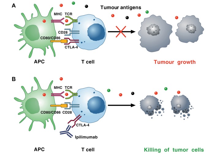
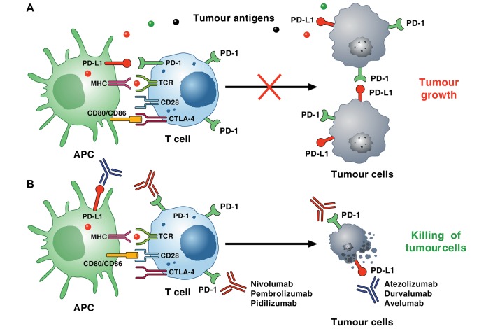
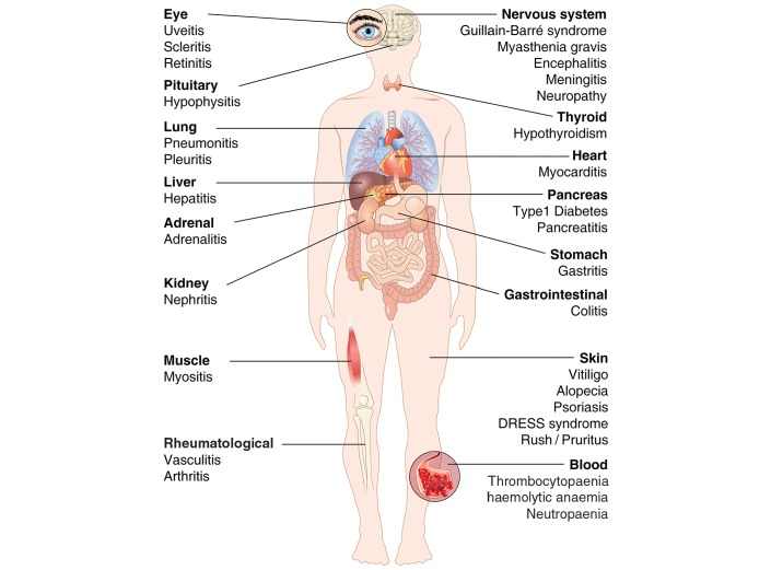
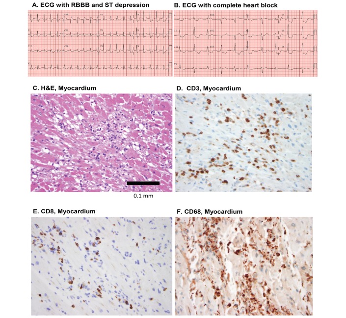
Cardiac toxicity after conventional antineoplastic drugs (eg, anthracyclines) has historically been a relevant issue. In addition, targeted therapies and biological molecules can also induce cardiotoxicity. Immune checkpoint inhibitors are a novel class of anticancer drugs, distinct from targeted or tumour type-specific therapies. Cancer immunotherapy with immune checkpoint blockers (ie, monoclonal antibodies targeting cytotoxic T lymphocyte-associated antigen 4 (CTLA-4), programmed cell death 1 (PD-1) and its ligand (PD-L1)) has revolutionised the management of a wide variety of malignancies endowed with poor prognosis. These inhibitors unleash antitumour immunity, mediate cancer regression and improve the survival in a percentage of patients with different types of malignancies, but can also produce a wide spectrum of immune-related adverse events. Interestingly, PD-1 and PD-L1 are expressed in rodent and human cardiomyocytes, and early animal studies have demonstrated that CTLA-4 and PD-1 deletion can cause autoimmune myocarditis. Cardiac toxicity has largely been underestimated in recent reviews of toxicity of checkpoint inhibitors, but during the last years several cases of myocarditis and fatal heart failure have been reported in patients treated with checkpoint inhibitors alone and in combination. Here we describe the mechanisms of the most prominent checkpoint inhibitors, specifically ipilimumab (anti-CTLA-4, the godfather of checkpoint inhibitors) patient and monoclonal antibodies targeting PD-1 (eg, nivolumab, pembrolizumab) and PD-L1 (eg, atezolizumab). We also discuss what is known and what needs to be done about cardiotoxicity of checkpoint inhibitors in patients with cancer. Severe cardiovascular effects associated with checkpoint blockade introduce important issues for oncologists, cardiologists and immunologists.

Keywords: CTLA-4; PD-1; PD-L1; cancer; cardiotoxicity; immune checkpoints; melanoma; myocarditis.


Conflict of interest statement

Competing interests: CGT received travel support from Alere.

Figures

Figure 1
Figure 1
Mechanism of CTLA-4-induced immunosuppression. (A) Cancer cells synthesise and release neoantigens (dots of different colours) that are captured by APCs. These cells present peptides in the context of MHC I molecules/TCRs on the surface of CD8+ cytotoxic T cells within lymph nodes. APCs can also present peptides bound to MHC II molecules to CD4+ T helper cells. T cell activation on TCR signalling requires costimulatory signals transmitted via CD28, which is activated by binding to CD80, and/or CD86, on the surface of APCs. Activated T cells upregulate CTLA-4, which competes with CD28 for binding to CD80 and/or CD86. The interaction of CTLA-4 with CD80 or CD86 results in inhibitory signalling promoting tumour growth. The immunosuppressive activity of CTLA-4 is mediated by downregulation of Th cells and enhancement of Treg cells. (B) CTLA-4 blockade by ipilimumab results in a broad enhancement of immune responses against neoantigen expressing tumour cells, which results in killing of tumour cells. APC, antigen presenting cell; CTLA-4, cytotoxic T lymphocyte-associated antigen 4; TCR, T cell receptor; Th cells, helper CD4+ T cells; Treg, regulatory T cell.
Figure 2
Figure 2
Mechanism of PD-1/PD-L1 pathway-induced immunosuppression within the tumour microenvironment. (A) Tumour neoantigens (dots of different colours) released by cancer cells are captured by APCs. These cells present peptides in the context of MHC molecules/TCRs on the surface of CD8+ cytotoxic T cells. PD-1 is induced on T cells on activation through the TCR and through several cytokines. Tumour cells and other cells in the tumour microenvironment (eg, endothelial cells, mast cells) can express high levels of PD-L1 and/or PD-L2 that binds to PD-1 on T cells, resulting in inhibitory checkpoint signalling that decreases cytotoxicity and leads to T cell exhaustion. Recent evidence suggests that murine and human cancer cell subpopulations can express PD-1 and promote tumour growth. (B) PD-1 blocking antibodies (nivolumab, pembrolizumab, pidilizumab and so on) inhibit the interaction of PD-1 with both PD-L1 and PD-L2, resulting in enhanced T cell cytotoxicity, TAM activity, increased cytokine production, and ultimately killing of tumour cells. PD-L1+ tumour cells can also induce T cell apoptosis, anergy, functional exhaustion and interleukin-10 production. Anti-PD-L1 antibodies (atezolizumab, durvalumab, avelumab) have similar effects, but only inhibit the interaction between PD-L1 and PD-1. PD-1, programmed cell death 1; PD-L1, programmed cell death ligand 1; TAM, tumour-associated macrophage; TCR, T cell receptor.
Figure 3
Figure 3
Some of the immune-related adverse effects (IRAEs) associated with checkpoints inhibitors in patients with cancer. DRESS, drug rash with eosinophilia and systemic symptoms.
Figure 4
Figure 4
ECG and histological findings of the heart in a 63-year-old man with metastatic melanoma who developed fulminant lymphocytic myocarditis following initial doses of nivolumab and ipilimumab and who developed complete heart block. Despite intense treatment (intravenous methylprednisolone 1 g/kg daily for 4 days plus infliximab 5 mg/kg), fatal complete heart block occurred. Initial right bundle branch block (RBBB) and ST depression (A) progressed rapidly to complete heart block and cardiac arrest (B). Autopsy showed lymphocytic infiltration in myocardium (C) comprised CD3+ T cells (D), many of which were CD8+ lymphocytes (E) and CD68+ macrophages (F) (adapted with permission from Johnson et al36).

References

    1. Bloom MW, Hamo CE, Cardinale D, et al. . Cancer therapy-related cardiac dysfunction and heart failure: part 1: definitions, pathophysiology, risk factors, and imaging. Circ Heart Fail 2016;9:e002661 10.1161/CIRCHEARTFAILURE.115.002661 - DOI - PMC - PubMed
    1. Hamo CE, Bloom MW, Cardinale D, et al. . Cancer therapy-related cardiac dysfunction and heart failure: part 2: prevention, treatment, guidelines, and future directions. Circ Heart Fail 2016;9:e002843 10.1161/CIRCHEARTFAILURE.115.002843 - DOI - PMC - PubMed
    1. Yeh ET, Bickford CL. Cardiovascular complications of cancer therapy: incidence, pathogenesis, diagnosis, and management. J Am Coll Cardiol 2009;53:2231–47. 10.1016/j.jacc.2009.02.050 - DOI - PubMed
    1. Tocchetti CG, Gallucci G, Coppola C, et al. . The emerging issue of cardiac dysfunction induced by antineoplastic angiogenesis inhibitors. Eur J Heart Fail 2013;15:482–9. 10.1093/eurjhf/hft008 - DOI - PubMed
    1. Suter TM, Ewer MS. Cancer drugs and the heart: importance and management. Eur Heart J 2013;34:1102–11. 10.1093/eurheartj/ehs181 - DOI - PubMed

LinkOut - more resources