

Case Reports
doi: 10.1155/2017/3191673.
Epub 2017 Sep 14.
The Foot That Broke Both Hips: A Case Report and Literature Review of Tumor-Induced Osteomalacia
Affiliations
- PMID: 29104808
- PMCID: PMC5618748
- DOI: 10.1155/2017/3191673
Item in Clipboard
Case Reports
The Foot That Broke Both Hips: A Case Report and Literature Review of Tumor-Induced Osteomalacia
Case Rep Rheumatol.
2017.
Abstract
Tumor-induced osteomalacia (TIO) is a rare paraneoplastic syndrome characterized by hypophosphatemia and clinical symptoms of osteomalacia. Only discussed as case reports, there is still limited knowledge of this condition as a potentially curable cause of osteomalacia among clinicians and pathologists. In this article, we present a case of tumor-induced osteomalacia in a 59-year-old gentleman followed by an up-to-date review of the existing literature on TIO.
Figures
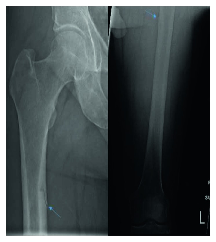
Radiographic image of both femurs with arrows pointing towards the fracture lines (the right femur on the left side and the left femur on the right side).
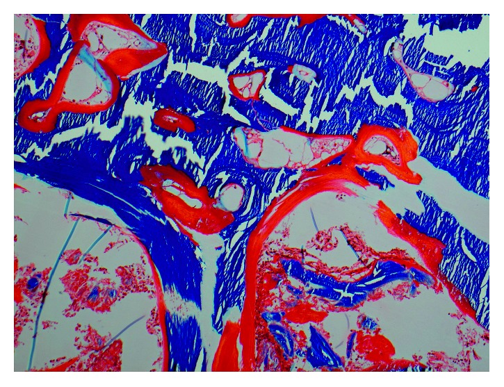
Plastic-embedded trichrome-stained pictures of the bone with 20x magnification. The trichrome staining shows wide seams of unmineralized osteoid (red) that covers virtually all trabecular surfaces, suggesting a severe mineralization defect.

Fluorescence microscopic pictures of the tetracycline-labeled bone with 20x magnification. A severe mineralization defect is confirmed by an unstained section of the bone under fluorescence microscopy looking for tetracycline labeling which reveals faint fluorescence with a blurred pattern and no evidence of the typical double labeling of the bone matrix.

Positron emission tomographic image of the lower extremities showcasing a fluorodeoxyglucose (FDG) avid spot on the plantar surface of the left foot (arrow).

Low-power microscopic image (2x) showing a well-delineated tumor consisting of variegated mesenchymal components rich in small vessels with focal myxoid stroma.

Low-power microscopic image (10x) showing a tumor consisting of prominent small vessels, myxoid stroma, and scattered osteoclast-type giant cells.

High-power microscopic image (40x) showing a tumor consisting of bland spindle cells sitting in slightly myxoid stroma with an osteoclast-type giant cell.
References
-
- Albright F., Butler A. M., Bloomberg E. Rickets resistant to vitamin D therapy. American Journal of Diseases of Children. 1937;54(3):529–547. doi: 10.1001/archpedi.1937.01980030073005. - DOI
Publication types
LinkOut - more resources
Full Text Sources
Other Literature Sources
